MORE POLLUTION MORE RESPIRATORY EMERGENCIES-DR.D.DK.JHA,M.D

November 7th, 2018

Air pollutin is a global emerging problem.

It affects all age groups but more affected age groups are children and elderly.

Bad air quality is known to exacerbate asthma in children and adults,and COPD in adults.

Now ,there is growing evidence that ,bad air quality may cause asthma in children and adults.

Particulate matters(PM) suspended in air may be inhaled and if the size is 2.5 micron or less(PM2.5),it may reach to the lower respiratory tract easily.

It may irritate the airways and cause asthma exacerbation.

If the airway is repeatedly irritated,it may become hyperreactive.

Other dangerous agent is ozone which adversly affects respiratory tract.

According to a study published in American Journal of Respiratory and Critical care medicine, people from areas of bad air quality depending on high ozone and PM2.5 level had high numbers of visits to the respiratory emergency department in comparision to the people from high air quality.

Data were analysed from 38.4 million visits to the department of respiratory emergency from 869 countries..Data represented 45% of US population including children,adults(18-65 years of age) and elderly beyond 65 years of age.Daily rates of respiratory emergency visits were 1.20 for all ages,1.90 for children,0.94 for adults and 1.37 for elderly over 65 years of age per 10000 population.The maximum 8 hour ozone per day varied from 8ppb to 34 ppb with a mean interquartile range of 16.54ppb.The level of PM2.5 ranged from 1.9mcg/m3 to 9.8mcg/m3 per day with an average of 5.3 mcg/m3. There was a positive association between respiratory emergency visits and ozone level.The rates of emergency visits were higher in children.

The study shows a causal relationship is likely between respiratory ailments and level of ozone and PM2.5 in ambient air

REFERENCES:


Strosnider H, Chang H, Darrow L, Liu Y, Vaidyanathan A, Strickland MJ. . Age-specific associations of ozone and PM2.5 with respiratory emergency department visits in the US [published October 2, 2018]. Am J Respir Crit Med. doi:10.1164/rccm.201806-1147OC


ANGIOEDEMA,DR.D.K.JHA,M.D

August 15th, 2018

Angioedema is characterised by sudden onset of swelling of eyelids,lips,tounge,genitals,dorsum of the hands and feet,which are visible on clinical examination and not clinically visible swelling of the gastrointestinal tract

Swelling of gastrointestinal tract present as pain abdomen of varying severity .Sometimes, acute severe pain like acute abdomen leading to unnecessary surgery.

Swelling involves the deeper subcutaneous layers.

There may be pain and itching but occasionally only pain is there.

It may take upto 72 hours for the resolution of sewlling.

It may be histamine related or non histamine related.

In histamine related angioedema,urticaria (wheals) will be present along with swelling

There may be a prodrome in the form of tightness or tingling of the area before swelling.

The most serious complication of angioedema is involvement of larynx in the form of laryngeal edema.

When the swelling involves the throat and uvula,it can readily be seen on clinical examination

It  may cause complete obstruction of airway needing intubation and sometimes tracheostomy to save the life.

Fortunately,life threatening episodes are rare.

Dental procedures,with injection of procaine into gum is a common precipitant but laryngeal edema may be spontaneous.

There is no response to epinephrine,antihistamine and corticosteroids on the swelling.


If there is only swelling without urticaria,it is not associated with histamine and it may of two types-HEREDITARY AND ACQUIRED ANGIOEDEMA.

HEREDITARY ANGIOEDEMA can be of three types;Type 1-reduced production of C1 inhibitor

Type 2-sufficient production but decreased function of C 1 inhibitor

Type 3-Estrogen dependent,due to mutation in factor xii seen mostly in women

ACQUIRED ANGIOEDEMA can also be of three types;Type 1-associated with reduced C 1esterase inhibitor

Type 2-Drug induced,most common offending drug is ACE inhibitor

Type 3-Histamine dependent regarded as urticaria without wheals

HEREDITARY ANGIOEDEMA is an autosomal dominant disease.

ACQUIRED ANGIOEDEMA may be seen in association with neoplasia,lymphoproliferative disorders and rarely infections.

In case of suspicion of hereditary angioedema type 1 and type 2, decreased levels of serum C 4,C 1inhibitor and decreased function of C 1 inhibitor should be assessed.

TREATMENT

Acute therapy is required during life threatening episodes,which is fortunately rare.

Specific treatment is concentrate of human C1 inhibitor ,the availability of which is limited

The approved drug is BERINERT,used as an intravenous formulation in the dose of 20 unit/kg

In case of non availability of this specific reatment,FFP(fresh frozen plasma) may be used which is least specific but widely available.

Bradykinin type 2 receptor antagonist ICATIBANT is approved for the age of 18 years and older

All treatmens are most effective when they are given early after onset of signs and symptoms

It may take 1-4 hours for the start of action after giving the drug.

Icatibant can be given in ACE inhibitor induced angioedema

Icatibant can be stored on room temperatue and can be given subcutaneously

Icatibant is licensed for home treatment in Europe.

CINRYZE-Purified C1-INH is approved for prophylaxix after acute attack in adolescents and older children

It is given in the dose of 1000 units intravenously twice a week

Danazol is another drug being used as short term prophylaxix ,but it is not given in children,

due to the side effects of premature closure of epiphysis

Danozol increases the production of C1 inhibitor in liver,

For histamine related angioedema, non sedating antihistamine is the drug of choice

In case of no response to usual  dose of nonsedating antihistamine ,the dose may be increased to 4 times

REFERENCES:

  • Borzova E and Gratton C ,Angioedema.In Zuberbier Tet al.Urticaria and AngioedemaBerlin:Springer Verlag 2010.122.
  • Bindslev-JensenC,CanonicaGV,Church mk,GimenzArnou AM et al. eaaci/ga2lan/edf/wao Guideline .Treatment of Urticaria .Allergy2010;64:1417-1426
  • Nelson Textbook of Pediatrics 20 e


OXYGEN IN ACUTELY ILL MAY BE HARMFUL-DR.D.K.JHA,M.D.

July 22nd, 2018

Oxygen therapy is a double edged sword in the care of acutely ill patients with hypoxemia and/or excessive use of accessory muscles of respiration.

It is a life saving therapy , but liberal use of it may increase mortality.

50%-80% of those receiving oxygen are exposed to excess oxygen and are at risk for hyperoxemia.

Liberal oxygen therapy may increase mortality at 30 days of hospitalization and at long term follow up according to the results of IOTA(improving oxygen therapy in acute- illness) ,study published in Lancet.

Derek K.Chu.MD and colleagues performed a systemic review and meta-analysis of studies identified using the cochrane central register of controlled trials,MEDLINE and WHO international clinical trials Registry.They included in their study,randomized clinical trials comparing coservative and liberal use of oxygen therapy in acutely ill adults.The study period was from inception to October 2017.

The study included 16037 patients in 25 randomized trials. The outcomes of  study were mortality and morbidity at longest follow up.Morbidity included hospital acquired pneuminia,hospital acquired infention of any type and length of hospital stay.Assessment of these outcomes wear done using random effects meta-analysis.

The relative risk(RR) of mortality was increased in the group with liberal use of oxygen(RR 1.21) for inhospital patients,30 day mortality(RR1.41) and longest follow up(1.10).The liberal use of oxygen also failed to improve the morbidity outcomes.

According to the suggestion of authors ,supplemental oxygen might be detrimental above a peripheral saturation range of  94%-96%.

Thus oxygen therapy should be used coservatively and judisiously to save life.Over enthusiastic treatment with oxygen may endanger life rather than saving it.

REFERENCE:Chu DK,Kim LH-Y,Young PJ,et al. Mortality and morbidity in acutely ill adults treated with liberal versus coservative oxygen therapy(IOTA):a systemic review and meta-analysis.Lancet.2018;391:1693-1705


WORLD ASTHMA DAY 2018 , PARENT AWARENESS/PUBLIC LECTURE BY PEDIATRIC RESPIRATORY SOCIETY DELHI

May 2nd, 2018

WORLD ASTHMA DAY IS OBSERVED ON FIRST TUESDAY OF EVERY MAY.

THEME OF THIS YEAR IS

NEVER TOO EARLY NEVER TOO LATE


STRERSS DURING ANTENATAL AND EARLY CHILDHOOD PERIOD MAY CAUSE ASTHMA,DR.D.K.JHA,M.D

March 30th, 2018

Asthma is the most common chronic disease in children.

Its incidence is increasing even in the era of advanced investigation facilities and treatment modalities’.

Most school days  of children are lost due to asthma morbidities.

Interaction between host and environment is necessary for the manifestations of disease in genetically susceptibles.

Stress is one of the aggravating factor for asthma flare ups.

It has long been assumed that the stress has a causative role in asthma and now a definite link has been found.

The exact mechanism for the causation of asthma by stress is still unclear.

There is more concerns by Researchers and clinicians about the developments during intrauterine life and during the first 2 years of life.

Stress during pregnancy affects the immune system,neuroendocrine system and antioxidant system.

The result is recurrent respiratory tract infections and recurrent wheezing and asthma in children born to such mothers having depression and anxiety during pregnancy.

Smejda and colleagues studied on 370 pairs of mother and child to know the correlation between maternal stress ,atopic dermatitis,asthma and recurrent respiratory infections.Those mothers were included who experienced stress for at least 1 month during pregnancy. The study was a part of Polish mother and child cohort,a large multicentre prospective cohort study established in 2007..The lead researcher was Katarzyna Smejda MD from the department of Pediatrics and Allergy ,Medical university of Lodz in Poland.The whole cohort comprises 1700 pairs of mother and child followed from pregnancy upto 2 years of age of the child. The ongoing cohort study is focussed on the child at the age of 7 years.

  •                One of the theories explaining the effects of maternal stress on respiratory health of children born to these mothers states that that ,there is increased levels of stress hormones particularly cortisol in mother who are in stress.
  • The cortisol crosses the placenta and binds to the endogenous and exogenous cortisol receptors of the fetuses and thus disturbs the hypothalmic-pituitory-adrenal axis.It also makes imbalance in the autonomic nervous system and immune system.The result is ,these children born of these mothers become susceptible to asthma and recurrent respiratory infections during early childhood.

Thakur and colleagues studied the effect of social discrimination on development of asthma in children aged 8-21 years.African American children who were discriminated socially had 78% more chance of developing asthma as compared to nondiscriminated(OR, 1.78; 95% CI,1.33-2.39). Children who felt discriminated socially also had poor asthma control even after good adherence to correct technique of asthma controller medication as compared to nondiscriminated choldren.(OR, 1.97; 95% CI, 1.42-2.76)

In conclusion,stress free environment during pregnancy and childhood is essential for a good respiratory health of children.

REFERENCES:

  • Rosa MJ, Lee AG, Wright RJ. Evidence establishing a link between prenatal and early-life stress and asthma development. Curr Opin Allergy Clin Immunol. 2018;18(2):148-158.
  • Busse WW, Kiecolt-Glaser JK, Coe C, Martin RJ, Weiss ST, Parker SR. NHLBI workshop summary: stress and asthma. Am J Respir Crit Care Med. 1995;151(1):249-252.
  • Smejda K, Polanska K, Merecz-Kot D, et al. Maternal stress during pregnancy and allergic diseases in children during the first year of life. Respir Care. 2018;63(1):70-76.
  • Thakur N, Barcelo NE, Borrell LN, et al. Perceived discrimination associated with asthma and related outcomes in minority youth: the GALA II and SAGE II studies. Chest. 2017;151(4):804-812.

EFFECTS OF ASTHMA AND MEDICATIONS ON FETUS DURING PREGNANCY,DR.D.K.JHA,M.D

January 26th, 2018
  • Asthma is the most common chronic respiratory disorders during pregnancy.
  • It affects 5%-8% pregnant women in United States.
  • Among the women suffering from asthma,almost one third experience exacerbation during second trimester of pregnancy.
  • Some women experience the symptoms of asthma first time during pregnancy.
  • It has been observed that,about one third of asthmatic women discontinue their asthma controller medications and reliever medications, during pregnancy fearing its adverse effects on fetus.

It has been observed that, there happens no chanage in FEV1,no admissible change in FEV1/FVC and  a slight increase in FVC ,during  normal pregnancy.There should be a cause of concern in case of any deviation seen in these observed parameters.

In normal pregnancy,there happens a change in cardiac output,stroke volume and heart rate.Women already diagnosed asthma may experience exacerbation due to these changes.Some women especially those who are genetically predisposed to develop asthma may experience the symptoms of asthma first time due to these changes during pregnancy.The enlarged gravid uterus my compress the inferior vena cava during third trimester of pregnancy,thereby decreasing cardiac output.This mechanical change along with some hormonal changes may exacerbate asthma and nonallergic rhinitis.

There is a strong correlation of asthma with obstetric and non obstetric comorbidities.Among obstetric ,the important ones are,preeclamsia,placenta previa,abruptio placenta eand obstetrical hemorrhage. There is higher rates of caesarean delivery in women with asthma.Among non obstetric comorbidities are increased rates of gestational Diabetes,increased rates of pulmonary embolism and increased frequency of respiratory infections including influenza.

Asthma exacerbation causes adverse effects on fetuses which includes,prematurity,low birth weight,fetal deaths(still birth) and some congenital anomalies in the form of cleft lip and cleft palate.Low birth weight has been seen more commonly in female while prematurity and still births have been seen more commonly in male fetuses.Children born to mothers having asthma have more chance of developing asthma.

Variuos research studies show that a good control of asthma symptoms and reducing or avoiding asthma exacerbation results in better outcome for mother and babies. There is a growing evidence in literatures that suggest that, inhalational corticosteroids(ICS) and beta-2 agonists(SABA), both are safe to be used during pregnancy for asthma management.Among ICS, the most studied is budesonide.ICS use during pregnancy improves FEV1 and reduces exacerbations.

The safety of biologicals in the form of IgE inhibitor(omalizumab) and anti IL-5(meplozumab and reslizumab),have not been established.However ,the woman already on these medications before becoming pregnant has not been seen to experience any adverse outcome.

Treating comorbidities is an important part to control asthma. If there is coexisting allergic or nonallergic rhinitis,it should be treated with intranasal sterioids.Coexisting GER should be treated with PPI . If there is coexisting anxiety and depression ,these should be addressed properly.

We should not forget to avoid allergen exposure in the form of house dust mite,animal danders,cockroaches,pollen and indoor molds. According to NAEP ,allergen impermeable bed coverings should be used,bed sheets should be washed weekly with water at or above 130 degree F,and humidity should be kept below 50%.

Good control of asthma by non pharmacologic and whenever needed pharmcologic measures gives better outcome for mothers and babies

REFERENCES:

1.Bonham CA, Patterson KC, Strek ME. Asthma outcomes and management during pregnancy [published online September 1, 2017]. Chest.pii: S0012-3692(17)31485-X. doi:10.1016/j.chest.2017.08.029
2.Meakin AS, Saif Z, Jones AR, Aviles PFV, Clifton VL. Review: placental adaptations to the presence of maternal asthma during pregnancy. Placenta. 2017;54:17-23.
3.Global Initiative for Asthma. Global strategy for asthma management and prevention, 2017. www.ginasthma.org Accessed October 3, 2017.
4.Liu X, Agerbo E, Schlünssen V, Wright RJ, Li J, Munk-Olsen T. Maternal asthma severity and control during pregnancy and risk of offspring asthma [published online June 28, 2017]. J Allergy Clin Immunol. 2017. pii: S0091-6749(17)30854-0. doi:10.1016/j.jaci.2017.05.016
5.National Heart, Lung, and Blood Institute; National Asthma Education and Prevention Program Asthma and Pregnancy Working Group. NAEPP expert panel report. Managing asthma during pregnancy: recommendations for pharmacologic treatment-2004 update. J Allergy Clin Immunol. 2005;115(1):34-46.
6.Bain E, Pierides KL, Clifton VL, et al. Interventions for managing asthma in pregnancy. Cochrane Database Syst Rev. 2014;10:CD010660. doi:10.1002/14651858.CD010660.pub2

NEW HOPE FOR BRONCHIOLITIS,DR.D.K.JHA,M.D

December 13th, 2017

Bronchiolitis is the most common resspiratory tract infection requiring hospital admission in infants.

The most common organism causing bronchiollitis is Respiratory syncytial virus(RSV).

Most children become infeccted with this virus by the second year of life.

Most of the infections are mild in nature and do not require any attention.

Few children acquire lower respiratory tract infection and some of them require hospitalization.

Other viruses causing bronchiolitis are Human Rhinovirus,Human Metapneumovirus,Bocavirus and many more

Children suffering from Rhinovirus bronchiolitis requiring hospital admission are more susceptible to develop asthma later in life.

Some children with bronchiolitis may be more susceptible to develop COPD later in adult life.

There is no definite treatment available to treat bronchiolitis.We are dependent only on supportive treatment in the form of maintenance of hydration and oxygenation of the child. Various inhalational medications are being used to treat this condition with variable results.Most frequently used inhalational medication is salbutamol.It is not clear that the child responding to salbutamol has a component of atopy and this is the reason for response. Other inhalational agents being used are adrenaline,normal saline,hypertonic saline,combination of salbutamol and ipratropium and inhalational corticosteroid. Some pediatrician use injectible corticosteroid to tide over the situation. Inspite of all the efforts,few childern face severe respiratory distress with prolonged course of hospital stay requiring intravenous fluid and oxygen.In rare instance, mechanical ventilation may be required to save the child.It is very important to note that, in children not requring hospital admission with normal feeding and activities,no treatment is required except saline nasal drop if there is nasal block and oral paracetamol if there is fever.

It has been proved that nitric oxide(NO) delivered at the rate of 160ppm ,effectively eliminate bacteria,virises,and fungi from respiratory tract.It is being used currently to treat serious lower respiratory tract infections  in adulds,not responding to standard of care treatment.It is also being used to treat pulmonary arterial hypertension9PAH0 in neonates.

AIT therapeutic inc,has a delivery system which affectively deliver NO at the rate of 160ppm.There has been phase 2 randomized double-blind controlled trial published in prestigious  Pediaric Pulmonology journal December 2017.In this trial, 43 infants between the age of 2-11 months suffering from moderatly severe bronchiolitis were enrolled. All received daily doses of inhalational nitric oxide 160ppm intermittently for 30 minutes ,five times a day for upto 5 days in addition to standard care in the form of IVF and Oxygen.There was no difference in side effects betwwen the study and control groups ,but length of hospital stay was significantly less in the study group.

Based on this result, Phase 3 trial(NO-BRO trial, nitric oxide in bronchiolitis)  has been initiated .It is a prospective  ranomized double- blind controlled trial. It will enroll 94 infants between the age of 0-12 months hospitalised with bronchiolitis. The study group will receive nitric oxide inhalation 160ppm for 30 minutes 5 times a day  for upto 5 days in addition to standard care and control group will receive only standard care in the form of IVF and Oxygen. The primary endpoint will be length of hospital stay and secondary endpoint will be time to achieve a score of 5 or less on modified Tal score  and time to achieve spo2 of 92% or more. The result is expected to come in the second quarter of 2018.

If rersults will be encouraging,it will be the boon for infants suffering from severe bronchiolitis and Pediatrician will breathe easily while facing the situation.

RFERENCES:

December 01, 2017 08:45 ET | Source: AIT Therapeutics, Inc.

Nitric Oxide Inhalations in Bronchiolitis: A Pilot, Randomized, Double-Blinded, Controlled Trial,Pediatric pulmonology Journal December 2017


Steven Lisi, Chief Executive Officer
AIT Therapeutics, Inc.
Steve@AIT-Pharm.com

Bob Yedid
LifeSci Advisors, LLC
Bob@LifeSciAdvisors.com
(646) 597 6989


VANISHING CARDIAC MURMUR ON STANDING POSITION IS NONPATHOLOGICAL, DR.D.K.JHA,M.D

November 26th, 2017

Cardiac murmur on routine clinical examination of children is not uncommon.

It affects approximately 65%-80% children in school going age.

It is the most disturbing clinical finding revealed to parents.

Clinicians are compelled to confirm its pathological nature by referring the child to cardiologists and getting a costly test done in the form of echocardiography.

Moreover,this echocardiography test is not available everywhere.

According to a study by Dr.Bruno Lefort and colleagues from Gatien de Clocheville Children hospital,Tours University hospital centre France, a cardiac murmur in children aged 2-18 years auscultated in supine position with no other cardiac sign and symptom and no family history of heart disease is almost always nonpathological, if the murmur vanishes when auscultated making the child in standing position.

In their study of 100 children with cardiac murmur in supine position that disappeard on standing position ,only 2 children were proved to have pathological murmur.

In the France study during 2014-2015,194 children referred consequitevely to cardiologist were first auscultated in supine position,then on  standing position and then echocardiography were done.The average age of the children was 6-7 years and most of them were boys.

30(15%) children were indentified as having pathological murmur by echocardiography in this study.

Murmur persisted on  standing position in 93% and decreased in intensity in 43%(p<0.001) children among pathological group. Among physiological group ,murmur persisted on standing position in 40% and decreased in intensity in 80%(p<0.001)

Abnormalities identified on decreasing order of incidence were Atrial septal defect(ASD),significant mitral regurgitation(MR),ventricular septal defect(VSD)and Aortic stenosis(AS)

The positive predictive value of excluding pathological murmur by its complete disappearance on standing position is 98%(95% confidence interval 93%-100%) with a specificity of 93%(95%confidence interval 78%-99%).Sensivity of this simple clinical examination was remarkably lower at 60%(95% confidence interval52%-67%)

Nonpathological or what we call an innocent cardiac murmur result from a normal blood flow through heart and great vessels. The cause of disappearnce of these physiological murmur on switching from supine to standing position is decreased venous return,left ventricular size and stroke volume.

Thus ,by the simple clinical examination of auscutating the heart in supine and then on standing position , a costly and not easily available echocardography may be avoided in most children with innocent cardiac murmur.

REFERENCES

Ann Fam Med. Published online November 13, 2017. Abstract

http://www.annfammed.org/content/15/6/523.full

NEW HOPE FOR PNEUMONIA DR.D.K.JHA M.D

November 11th, 2017

Pneumonia is responsible for highest number of death in children below 5 years of age worldwide.

The highest number of pneumonia cases in children are found in India.

There are numerous causes of pneumonia including viruses,bacteria,fungi among infectious causes.

Streptococcus peumoniae and Haemophilus influenzae type b(HIB) account for 60% of cases of community acquired pneumonia in Indian children below 5 years of age .

These community acquired bacterial pneumonia are treated by antibacterial agents(ANTIBIOTICS) as a mainstay of treatment.

There is an increasing incidence of resistance of these organisms to existing antibiotics.

Moreover,there are very few antibiotics in the pipeline research to fight these resistant bacteria.

There is urgent need of a new antibiotic to fight resistant community acquired bacterial pneumonia.

World health organisation(WHO) has oversimplified the definition of pneumonia so that no case of pneumonia should be misssed even by ground level health workers.

According to WHO

NO tachypnea=No peumonia

Tachypnea=Pneumonia

Tachypnea with chest retraction=Severe pneumonia

Tachypnea with chest retraction with cyanosis/lethargy/poor feeding=very severe pneumonia

According to the definition and categorization by WHO, pneumonia is overdiagnosed as these findings may be present in children suffering from respiratory diseases other than pneumonia like bronchiolitis and asthma.

Pneumonia in children is diagnosed by a combination of clinical features and investigations.

CLINICAL FEATURES: Fever

Tachypnea:(Respiratory rate>60/minutes below 2 months of age,>50/minute between 2-12 months of age,>40/minute between 1-5 years of age and more than 30/minute between 6-8 years of age)

Tachycardia:(Pulse rate>160/minute between 2-12 months of age,>120/minute between 1-2 years of age and >110 between 2-8 years of age

Chest retraction In the form of subcostal,intercostal and suprasternal retraction

Auscultatory finding: Diminished intensity of breath sound over affected area and deep inspiratory crackles are indication of consolidation in lung parenchyma which is pathognomonic of pneumonia.Bronchial breath sound and wheezes may be heard.

In severe pneomonia, clinical cyanosis may be visible or hypoxia may be detected by pulse oximetry.

INVESTIGATION:Complete blood count may show leucocytosis with predominance of polymorphoneuclear cells.

CRP is significant only if it more than 20mg/dl and blood culture is positive in only 15-20% cases

CHEST X-RAY : it may show nonhomogenous opacity in the lung field indicative of consolidation.Air bronchogram if visible is pathognomonic of consolidation.If opacity is involving a lobe ,it is lobar pneumonia and if the opacity is in bronchial distribution bilaterally, it is bronchopneumonia. Pneomatocele may be visible in Staph Pneumonia.

TREATMENT;The mainstay of treatment of bacterial pneumonia is antibiotics with or without supportive care in the form of intravenous fliud and oxygen if needed.

Oral amoxycillin is the drug of choice in OPD cases.If resistance to it is suspected the doses may be  doubled or a combination of amoxycillin and clavulanic acid may be given.Alternative agents are cefuroxime or cefprozil. Azithromycin should be given only when Mycoplasma or Chlamydia pneumonia are suspected.

In admitted patient the drug of choice is intravenous third generation  cephalosporin.If Staph Pneumonia is suspected Vancomycin or Clindamycin should be given.It should be for 3 days after the child becomes afebrile or for 10 days whichever is later.

There is a new hope for the growing resistance of bacteria to commonly used antibiotics.

Lefamulin belongs to a new class of antibiotics called Pleuromutilins.

Is has the same clinical profile as moxifloxacin which is also used to treat resistant tuberculosis apart from community acquired bacterial Pneumonia.Similar rates of adverse affects has been seen in both the drugs.The efficacy to kill bacteria is also the same.Lefamulin acts by binding to a specific site of bacterial ribosome responsible for protein synthesis.Retapumilin was the first drug of Pleuromutilin group ,approved for human use.This drug Retapumilin is approved for topical use.

Lefamulin has been tried successfully and it is in the final stage of trial . Probably it will hit the the market in the later half of 2018 if approved by FDA.If so,it will be the first drug approved for oral or intravenous use from Pleuromutilin group.Once it will come in the market it may help in treating resistant community acquired bacterial pneumonia and it may spare the drug moxifloxacillin for its use in resistant Tuberculosis as well.

REFERENCES:1.WHO Library Cataloguing-in-Publication Data
Revised WHO classification and treatment of pneumonia in children at
health facilities: evidence summaries.
1.Pneumonia – drug therapy. 2.Child. 3.Health Facilities. 4.Guideline.
I.World Health Organization.
ISBN 978 92 4 150781 3 (NLM classification: WA 320)

2.Expert panel report 3,guidelines for the diagnosis and management of asthma,NIH publicationNo.07-4051,Bethesda ,MD, 2007.U.S Dpartment of health and human services;National institute of health,National heart,,lung and blood institute,National Ashma Education and Prevention Program.

3.Nabriva’s Pneumonia Drug Succeeds in Late-stage Trial – Medscape – Sep 18, 2017.

4. Nelson Text book of Pediatrics ,edition-20


Newer Inhalational Control Therapy for Asthma

November 5th, 2017

Tiotropium has been studied successfully for control of asthma in children

NEWER DRUG FOR ASTHMA CONTROL -DR.D.K.JHA,M.D.

November 5th, 2017

Asthma is the most common chronic respiratory disorder of children.

It is a pleomorphic disorder characterized by hypersensitivity induced bronchospasm,airway inflammation and variable airway obstruction.

More than 100 genes have been recognised which predispose a child to develop asthma,the most important is 17Q21

Most of the asthmatic children respond very well to inhaled corticosteroid(ICS) alone or in combination with long acting beta agonist(LABA) for the control of asthma symptoms and disabilities.Some children require LTRA in the form of monteleukast as an add on therapy to ICS and LABA for the control of asthma.

Rarely some children do not respond in spite of correct technique of inhalational medications , good compliance,allergen avoidance and comorbidities well addressed.

There is practically no drug available for these children below 12 years of age.

It has been well established that interleukin-4(IL-4) and interleukin-13(IL-13) are responsible for intiation of airway inflammation in asthmatic airways.So,some biological agents are being tried to block these inflammatory initiators.

A new biologic agent DUPILUMAB which blocks IL-4 and 1L-13 has shown promosing results to control severe  asthma in adults.

A study in this regard has been done by Jonathan Corren MD,David Geffen School of medicine,university of California,Los Angeles.According to him, the severity of asthma is best assessed by number of exacerbations in a span of time.He has studied on465,227,122,62 patients with asthma  with 1 0r more,2 or more,3 or more and 4 or more exacerbations respectively in one year.He had given 200mg to 300mg DUPILUMAB at an interval of 2 weeks in addition to ICS and LABA for 24 weeks.

The additioon of Dupilumab has improved the quality of life as assessed by Asthma quality of life questionnaire (AQLQ) and Asthma control questionnaire(ACQ 5,5items)..The impact was seen regardless of eosinophil counts.There was no difference in events of adverse effects among subgroups.Patients with greater number of exacerbations benefitted more.

This study warrants a multicentre study in children so that the children with very poorly controlled asthma even on appropriate therapy may benefit.

Lancet. 2016;388:31-44


DENGUE FEVER LEADING TO HLH ,A POTENTIALLY FATAL CONDITION,D.K. JHA,M.D,CASE REPORT

February 10th, 2017

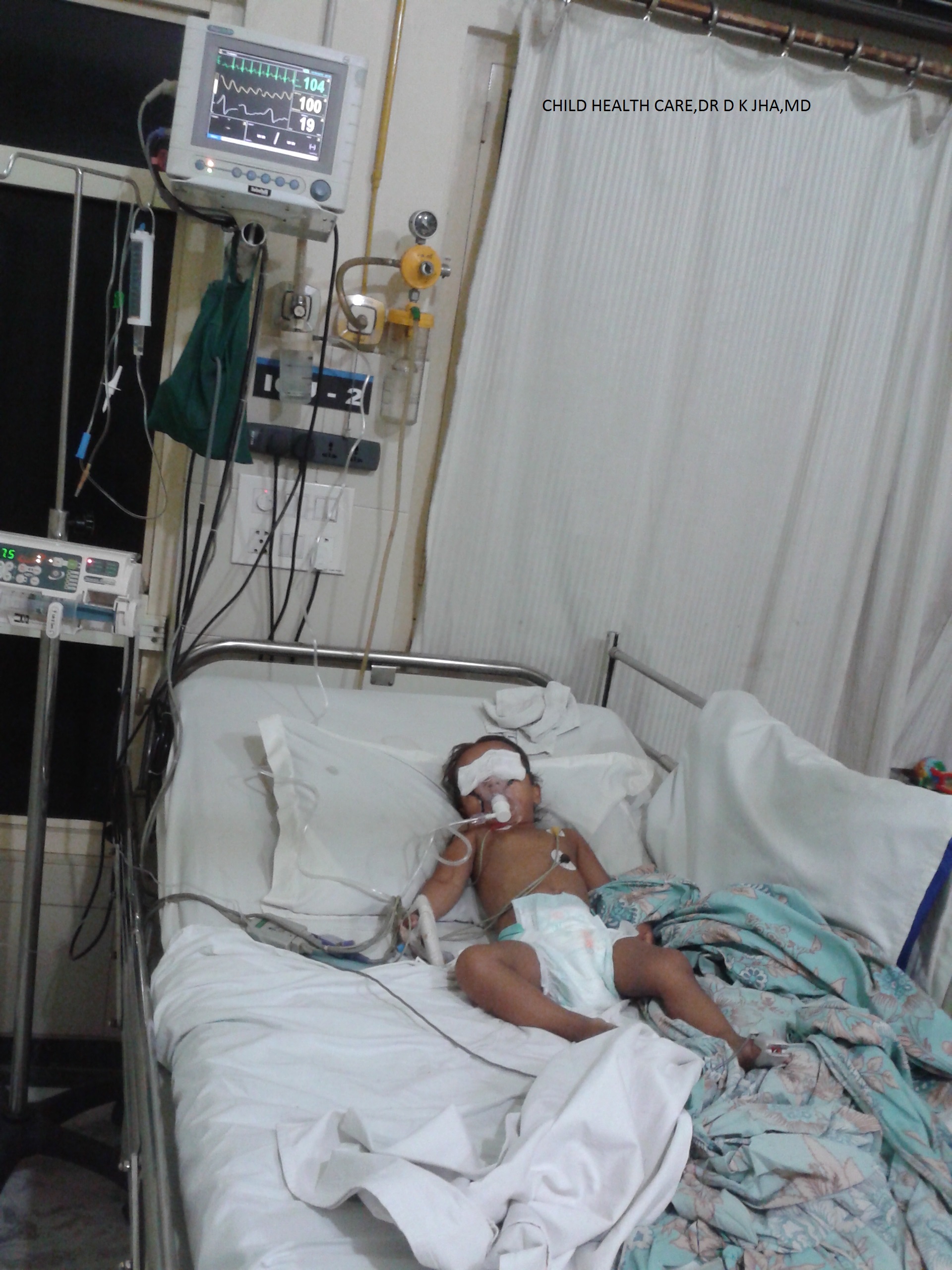
Faizan is a 12 years male child who was referred to us due to unmanageable fever with features of shock.

At our centre there was no fever ,the child had not passed urine in last 12 ours,pulse was not palpable and

blood pressure was 80/60mmhg,tongue was dry but not coated,chest clear on auscultation,CNS -NAD,P/A Liver 2cm and Spleen just palbable.


On exploring the history,the child was having fever off and on for last 6 weeks being treated by local doctor,with high fever for last 4 days ,pain abdomen for last 3 days and vomiting sensation but no vomiting with generalised body pain for last 2 days.

There was no history of cough,cold,loose stools,or burning urination.No H/O repeated ear discharge,repeated skin rashes,repeated headache or abnormal body movement with loss of conciousness.

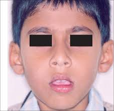
he was involved in wrestling competition for last 1-2 years and I suspect he was taking some muscle making drugs in the form of steroids.His weight was 50 kg and was looking like an adult.

He was given intravenous normal saline boluses to treat the shock and blood was withdrawn for investigations.

He recovered from shock in next 24 hours without catacholamimnes,started to pass urine normally ,pulse became palpable with a rate of 72/m and Blood pressure 1oo/70mmhg then 110/80mmhg

He was carrying some investigations reports which was as under

Hb 17gm with hematocrit 51%

TLC 1100 with 80%polymorph

Platelet count 24000/cmm

Malaria antigen negative

We thought,we are dealing with dengue shock syndrome,then reports from our centre came which was as under

After 12 hours of starting treatment blood picture was as below

Hb 14gm/dl with hematicrit 42%,TLC 9000,with 80%polymorph

Platelet 32000/cmm

He was on treatment with IVF ,iv ondensatron and iv pantoprazole

After 7 hours blood count was repeated and report was as below

Hb 11gm,TLC 8000 with 80% polymorph

Platelet count 32000/cmm

At this time other reports came which was as under

S.BIL 0.9mg,SGPT 1300IU,SGOT1350IU

S.SODIUM 122MEQ/L,S.POTASSIUM 4.5MEQ/L,BLOOD UREA 56mh/dl.S.CREATININE 1.3mg/dl

S,\.ALBUMIN 3.4GM,S.GLOBULIN 4.2GM,A/G 0.8

SERUM WIDAL 1:160

DENGUE IGM NEGATIVE

DENGUE IGG POSITIVE

MALARIA ANTIGEN NEGATIVE
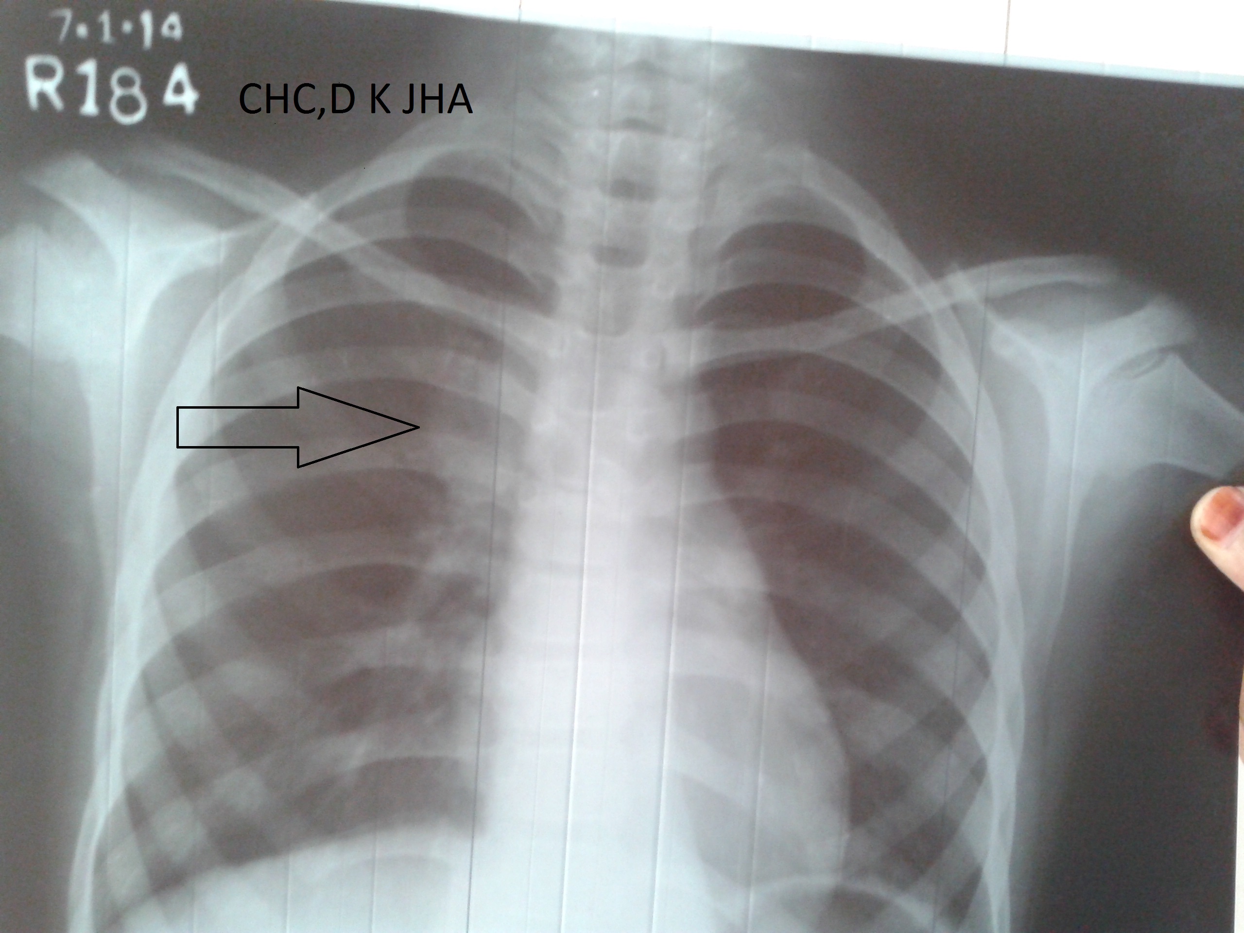
CHEST X-RAY NORMAL

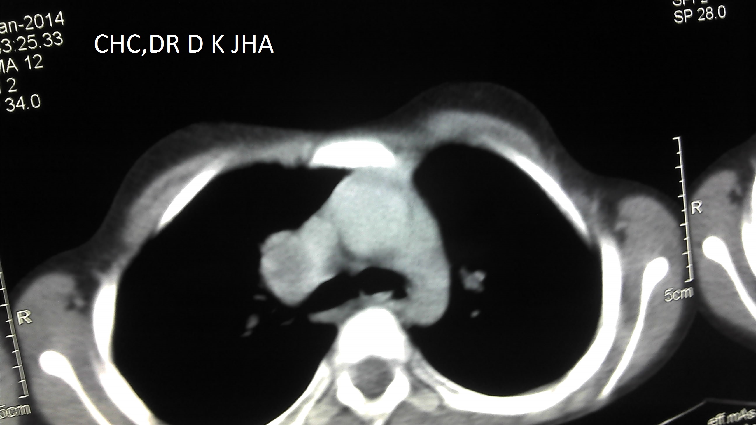
USG ABDOMEN bilateral mild pleural effusion,withj mild ascites with edematous gall bladder wall

so, these were consistent with viral infection,probably with dengue virus with salmonella infection

We started iv ceftriaxone

After 15 hours blood picture was as below

Hb 8gm with hematocrit 25%,TLC 4000,

Platelet 22000/cmm

AT this I requested some special investigation .and previous investigation repeated after 36 hours and report was as below

SGPT 3500IU,SGOT 3700IU,

S.SODIUM 129MEQ/L,S.POTALOODSSIUM4.5MEQ/LS.CREATININE 1MG/DL,BLOOD UREA 34MG/DL

S.ALBUMIN 2.4GM.S.GLOBULIN 3GM/DL,A/G 0.8

Platelet count 22000/cmm

Now the child developed fever which was recorded 101degreeF

At this time 6 units platelets was transfused after giving oral paracetamol ,when fever came down.

Child was feeling better and started to take orally

There was no bleeding from any obvious site throught the illness

Platelet count was repeated after 2 hours of transfusion

and it was still 22000/cmm

NOW SPECIAL REPORTS CAME WHICH WAS AS UNDER

S.TRIGLYCERIDE 165MG/DL

S.CHOLESTEROL 126MG/DL

S.FERRITIN 12000ng/ml

At this time the child was diagnosed as a case of

HEMOPHAGOCYTIC LYMPHOHISTIOCYTOSIS (HLH) probably due to Dengue virus infection.

HLH is a condition of uncontrolled activation of macrophages and lymphocytes with storming release of cytokines leading to multiple organ damages and consequent clinical manifestations.

It is of two types:

PRIMARY HLH

SECONDARY HLH

Primary HLH is due to congenital defects leading to uncontrolled activation of macrophage and lYmphocytes,whereas secondary HLH is due to various infective and non infective conditions,leading to uncontrolled activation of macrophages and lymphocytes.

PATHOPHYSIOLOGY

Macrophages which are derived from monocyets act as a presenter of antigens to the     lymphocytes .

A subset of lymphocytes called NK cells (Natural killer cells) and CTL (cytotoxic T cells) are involved in  killing and washing out activated macrophages from blood cells pool when their work is over and are no more needed.If  NK cells and or CTL becomes less effective or  ineffective in number and function, either due to congenital or aquired cause,there will be uncontrolled increase in number of activated macrophages ,uncontrolled activation of lymphocytes and histiocytes and storm like release of cytokines.This is the reason,this condition is also called as macrophage activation syndrome.

Cytokines released are interleukin1,interleukon6,interleukin10,gamma interferon and tumor necrosis factor alpha with some others interleukin 2 soluble receptor(CD25)

These cytokines creats aggressive inflammation throughout the body and there is engulfment of blood cells like RBC,WBC and Platelets by activated macrophage,lymphocyte and histiocytes .This is why, the condition is known as hemophagocytic lymphohistiocytosis(HLH).This is condition of aggressive but ineffective immune response which ultimately damages multiple host organs.

Hemophagocytosed macrophages demonstration in bone marrow is not necesary to diagnose this condition and moreover it is not pathognomonic.

ETIOLOGY:

PRIMARY HLH : There are 5 types of genetic mutations known till date .PRF and SAP mutations are important among them.

SECONDARY HLH:Activated by infective and noninfective conditions.

Among infections, the common causes which may trigger HLH often in the setting of primary or secondary immunodeficiency are

Viral-Epstein-Barr virus,CMV,Dengue virus

Bacterial-Enteri Gram negative rods ,Staphylococcus,Streptococcus,Mycobacterium tuberculosis

Fungal-candida albicans

Parasitic-Leishmania donovani,Plasmodium

Ricketsial-coxiella burnetti

NON INFECTIVE CONDITIONS:

Primary immunodeficiency diseases,secondary immunodeficiency due to any cause, some malignancies like leukemia and lymphoma, autoimmune diseases like systemic onset juvenile idiopathic arthritis,systemic lupus erythmatosus

CLINICAL FEATURES:The primary HLH usually present in infancy but may present later on at any age.The secondary HLH depends on the age of onset of inciting diseases.The disease present with fever,malaise,irritability,restlessness,vomiting and some times with respiratory distress..On clinical examination,one may find pallor,icterus,generalised lymphadenopathy,splenomegaly,hepatosplenomegaly,generalised body rashes which may be maculopapular pruritic or morbilliform rashes,symptoms of CNS involvement similar to meningitis or acute demyelinating encephalomyelitis may be seen.

ON INVESTIGATION;BLOOD– common findings are anemia,pancytopania,hyperbilirubinemia,increased transaminases,hypoalbuminemia,persistent hyponatremia,hyperferritinemia usually >10000ng/ml,hypofibrogenemia,hypertriglyceridemia and decreased ESR

ON RADIOIMAGING:CXR may reveal bilateral pleural effusion and USG abdomen may reveal edematous gall bladder with its wall thickening and ascites.MRI brain may show hyperintensities in gray and white matter along with supratentorial and infratentorial regions.

DIAGNOSIS:It is established by either demonstration of genetic mutations consistent with HLH which is available at few selected centres worldwide or fulfilling 5 out of following 8 criteria

1.Fever of more than 5-7 days

2.Splenomegaly:palpable spleen in the left subcostal region

3.cytopenia(at least 2 cell line affected-Hb equal to or<9g/dl : equal to or <10gm/dl for neonates,platelet<100000/cc,neutrophil<1000/cc

4.Hypertriglyceridemia(equal to or>265mg/dl) and or hypofibrogenemia(equal to or<150mg/dl)

5.Hyperferritinemia(serum ferritin equal to or more than 500ng/ml)

6.Increased level of CD 25 in serum(equal to or more than 2400U/ml)

7.Decreased or absent NKcell (Natural Killer cell) activities)

8.Demonstration of hemophagocytosis in bone marrow,spleen or lymph nodes

Many times, less than 5 criteria are met and even then the diagnosis is made .Moreover,in upto 70% cases, the initial bone marrow examination does not show hemophagocytosis ,which may be found later on.

TREATMENT:For primary HLH ,when genetic mutation is established the current recommendations are

Etopocyde

corticosteroids

Intrathecal methotrexate

These should be given even in presence of pancytopenia.These treatments are given to calm down the hyperimmunologic response so as to lessen the organ damage.The definitive treatment is Allogenic Stem cell transplantation.

For secondary HLH , treatment is aimed at treating and controlling the underlying infective or noninfective conditions along with supportive care.If there is any evidence of iatrogenic immunosuppression,the immunosuppressive agents are withdrawn and underlying infections are treated along with supportive care. Most of the time, for non infective causes or no known or untreatable infections ,Etoposide remains the drug of choice.Other agents are interferon and intravenous immunoglobulin.Etoposide has cytotoxic effect on macrophages ,thus decreases cytokine production and hemophagocytosis,limiting the organ damages.

PROGNOSIS:For primary HLH the prognosis is invariably poor with certain fatality. Even after appropriate treatment the disease may be suppressed to again flare up after some time and death occurs. The only cure is Allogenic stem cell transplantation which is being done at certain centres in the world including India.

For secondary HLH ,the prognosis may be excellent if infection is the causative factor,it is timely diagnosed and appropriatley treated if treatable.. For non infective causative factors the prognosis is poor with very high mortality.

REFERENCES:

1.Stephan Ladisch,Hemophagocytic Lymphohistiocytosis volume 3,Nelson Textbook of Peditrics,20e,2016,2488-2489

2.[Guideline] Henter JI, Elinder G, Ost A. Diagnostic guidelines for hemophagocytic lymphohistiocytosis. The FHL Study Group of the Histiocyte Society. Semin Oncol. 1991 Feb. 18(1):29-33. [Medline].

3.Pal P, Giri PP, Ramanan AV. Dengue associated hemophagocytic lymphohistiocytosis: a case series. Indian Pediatr. 2014 Jun. 51(6):496-7. [Medline].

4.Gupta S, Weitzman S. Primary and secondary hemophagocytic lymphohistiocytosis: clinical features, pathogenesis and therapy. Expert Rev Clin Immunol. 2010 Jan. 6(1):137-54. [Medline].

5.http://www.uptodate.com/contents/clinical-features-and-diagnosis-of-hemophagocytic-lymphohistiocytosis/abstract/20

UNUSUAL MODE OF PRESENTATION OF INTRATHORACIC TUBERCULOSIS,DK JHA ,MD

July 19th, 2016

Sania is 8 years ,female child,came to our OPD,with complaint of pain abdomen for 3-4 days.

There was no diurnal variation in pain and no relation to food intake or fasting.

The pain was not associated with vomiting or any change in bowel habit.There was no fever and no change in color or frequency of urine or burning sensation while passing urine.

The pain was not severe enough to interrupt her normal daily routine and she was attending her school  as usual with no medication.

On examination,her anthropometry was appropriate for the age,general examination did not reveal any deviation from normal finding.Abdominal examination was normal,and other systemic examination was non cotributotry.

She was prescribed antihelminth in the form of Albendazole   tablet, in view of high incidence of worm infestations in the area from she is coming,and pain abdomen is the usual symotom of worm infestations.

After two weeks ,she again came to OPD with pain abdomen and this time she was crying with pain since last night with vomiting tendency but no vomiting.She was admitted in general ward(Pediatrics).Symtomatic treatment was given and investigation was done to search the cause of pain abdomen. Urine and stool examination was normal.Ultrasound of abdomen showed mild enlargement of left kidney but KFT was normal.CBC did not show any marker of infection.The child was discharged as she improved clinically. She was adsviced to visit OPD with repeat ultrasound examination of abdomen after 2 weeks . She visited OPD with USG abdomen report ,which was normal this time. But  the pain abdomen was persisting which was not interfering with her normal routine. After one week she came in OPD with complaint of getting difficulty in standing from sitting position.On examination there was no vertebral tenterness but the difficulty was confirmed.X-Ray thoracolumbar spine was done which was normal. Chest X-Ray was done to search the cause which revealed right hilar lymphadenopathy . Then Montoux test was done which was 22mm.GA for AFB was negative and GeneXpert test of GA did not show AFB.There was no contact history of tuberculosis. Then CECT Chest was done with contrast which revealed right hilar lymphadenopathy with central necrosis and prevertebral collection in thoracic region. The child was diagnosed as intrathoracic tuberculosis and ATT was given under CAT I and the child was responding to treatment.After 3 months of treatment the child again developed pain abdomen with vomiting.She was admitted again as oral intake was very poor.It was thought that there may be drug induced hepatitis as icterus was present and liver was enlarged and tender.Laboratory report showed s.bil 8.5 mg/dl with direct bilirubin 5mg/dl.SGPT was 1300IU/L and SGOT was 1050 IU/L.Blood was sent for viral markers and ATT was withheld.The child was improving symptomatically.The viral marker report showed Hepatitis E.Child was improved within 10 days with normal bilirubin and liver enzymes. ATT was resumed at the time of discharged and on follow up child was doing well with continuation phase of ATT which was prolonged for 2 weeks more.On completion of ATT,the child was asyptomatic,gained 3 kg weight(from 22kg to 25 kg) and repeat CXR was normal. The child is doing well in follow up for last 2 years.20140819_10355620140819_1036262014-01-30_11-11-04_6662014-01-30_11-09-41_53820140819_103656

SOME FACTS ABOUT TUBERCULOSIS:

  • The term tuberculosis was given by Schonlein in 19th century.
  • This term is derived from the English word tubercle which means lesion of consumption.
  • The causative organism is mycobacterium tuberculosis which is a weakly gram positive curved rod.
  • All mycobacteria form stable mycolate complexes with arylmethane dye which resist decolouration with etanol ,or any acid,so these are also known as acid fast bacilli-AFB
  • Thoracic tuberculosis in children is hilar or mediastinal  lymphadenitis.
  • Most common type of extrapulmonary tuberculosis in children is cervical lymphadenitis.
  • Common lymph nodes involved are tonsillar,submandibular,anterior cervical and supraclavicular.
  • These lymph nodes enlargement should be differentiated from causes like cat scratch disease,tularemia,Brecellosis and Non mycobacterial tuberculous infections.
  • Tuberculous lymphadenitis is also called Scrofula which is more common in children than adults.
  • Even after proper treatment the size of lymph node may remain enlarged for months or even years.
  • person having rare genetic defect of cell mediated immunity are more susceptible to this disease.
  •  like interleukin 12 receptor B1 deficiency and complete or partial gamma interferon receptor 1 chain deficiecy are particularly susciptable for tuberculous disease.
  • Some group are specially susciptable to this disease known as Mendelian susceptibility to mycobacterial disease-MSMD
  • Person having certain HLA type and containing Bcg gene are predisposed for this disease.
  • This is the second most common killer infection of the world second only to HIV
  • Almost one third population(2.5 billion) is infected with this disease causing organism Mycobacterium tuberculosis.
  • Once infected the lifetime risk for converting into disease for an adult is 5%while in infants it is about 40% within 1 year and in children it is about 10%.
  • Children in the age group of 5-15 years are relatively less sususceptible for the disease.
  • Inhalation of 5-200 bacilli are needed to cause infection but even a single baicillus can cause the infection.
  • one infective droplet nucleus in air contains 1-10 bacilli and its size is approximately1- 5 micron which is easily respirable and remain suspended in air for long time.
  • A tuberculous nodular lesion contains 100-10000 bacilli while a cavity contains 10 million-1billion bacilli making it highly infective.
  • Infection is more common at crowded place where air circulation is low.
  • Infections through fomites have not been reported till now.
  • Infections like measles,varicella and pertussis make the latent tuberculous infections,active.
  • Children acquire the infection from adult suffering from pulmonary tuberculosis,while children to children transmission is rare because of very low bacterial load in children.
  • Most common type of tuberculosis in children is intrathoracic tuberculosis.
  • Most common type of intrathoracic tuberculosis in children is right hilar lymphadepathy then right paratracheal lymphadenopathy.
  • Most common symptom of thoracic tuberculosis in infants are non productive cough and mild dyspnea.
  • Cough may persists for  few months in children even after proper treatment of reavtivation pulmonary tuberculosis.
  • Fever persists for months after proper treatment of miliary tuberculosis and for weeks after proper treatment of tuberculous pleural effusion.
  • Most of the adult patients of pulmonary tuberculosis become non infectios after 2 weeks of proper treatment but some may transmit disease for several weeks even on treatment.
  • Once a person suffers from disease, it usually provides life time protection for subsequent disease, but second time disease has been reported in same individual.

SOME IMPORTANT TERMS USED IN CONNECTION TO TUBERCULOSIS

Exposure: This term is used when there is a definite history of contact with a person having pulmonary tuberculosis but no evidence of infection in the form of TST positive or IGRA positive

INFECTION: This term is used when there is evidence of infection in the form of positive TST (Mantoux test)or positive IGRA (Quantiferon gold)but no symptom ,no sign on clinical  examination and no radiological abnormality.This is also called LTBI (Latent Tuberculous bacilli infection)

TST(Mantoux test)becomes positive after 3-12 weeks of exposure. So exposed person may be having infection even if Mantoux test or IGRA test is negative within this period.

Mantoux test is said to be positive when induration is 10mm or more.In some countries the cut off for BCG vaccinated child is 15mm.

In HIV positive children and children with severe acute malnutrition ,induration of 5 mm or more is considered positive.

IGRA(Quatiferon gold) test is not reliable below 4 years of age.

MONTOUX POSITIVE OR IGRA POSITIVE STATUS INDICATES ONLY INFECTION ,NOT THE DISEASE,WHEREASE THE NEGATIVE MONTOUX TEST DOES NOT RULE OUT THE DISEASE.

DISEASE:When there are symptoms,signs and radiological changes associated with tuberculous infection,it is called tuberculous disease and AFB may be demonstrated at this stage by various diagnostic modalities.

Only disease state is treated in India because of large prevalence of infection in this country.In western countries LTBI is also treated.

VARIOUS FORMS OF TUBERCULOSIS IN CHILDREN:

Primary pulmonary tuberculosis: This is the most common type of tuberculosis in children. In this category the bacilli enter the lung through inhalation and get lodged into lung lobe at a site just below pleura.All lung lobes are equally affected. Macrophages engulf the bacilli and cytokines released by lymphocytes attack the bacilli to kill them.There is tissue reaction and formation of epitheloid granuloma(ghons focus) and caseation at the centre of granuloma.Some bacilli which escape killing travel through lymphatics to the regional lymph nodes at the hilum.There occurs tissue reaction in the lymph nodes and its size enlarges.The enlarged lumph node along with primary focus of infection is collectively called PRIMARY COMPLEX.The enlarged lymph nodes may press the bronchus.Partial compression of bronchus causes hyperinflation of lung lobe by ball valve mechnism.When there is complete obstruction there becomes collapse of the segmental.Sometimes the lymph node erodes the bronchus and discharge caseous material into bronchus which travels to the lobe causing consolidation.Then it gives the picture of segmental collapse consolidation on chest Xray.It is also known as segmental lesion.Sometimes caseous material blocks the bronchus and causes complete collapse .Sometimes the primary infection is progressively destructive causing liquifaction of pulmonary parenchyma and then a thin walled cavity is formed. In this form of tuberculosis bullous lesion may develop which may rupture to form pneumothorax.The pathognomonic finding  of this  form of tubercolosis on chest Xray is relatively large size of lymph node as compared to parenchymal lesion.

Signs and symptoms are usually meagre at this stage of tuberculosis.Diagnosis is suspected by history of contact with not gaining weight or failure to thrive in an appropriate clinical setting.

Primary Progressive Pulmonary Tuberculosis: In this form, tuberculosis presents as pneumonia which may be expansile pneumonia.Signs and symptoms are prominent like bacterial pneumonia .Common symptoms are weight loss and and night sweats along with high  fever and severe bouts of cough,which points towards diagnosis of Tuberculous pneumonia.,There may be lobar consolidation which may progress with caseation at the centre.It may erode the bronchus and spreads through bronchi to different areas of lungs. There may be formation of cavities when there is liquifaction of the lesion.Cavities may contain numerous bacilli which becomes dangerous to the surrounding population when there is vigourous cough.

Reactivation Tuberculosis:It occurs 1 year after primary healed tuberculous   infection, when the primary sites , commonly the original primary focus,lymph node or   lung apex  which were seeded during primary tuberculosis reactivates due to depressed immunity of individual.Pulmonary apical seedings are known as Simons foci.It is seen usually in adults,but children rarely suffer from this form, when children acquire primary infection after 7 years of age. In this form of tuberculosis ,seen usually in adolescents, symptoms are there in the form of fever,loss of appetite,loss of weight,night sweats,cough which is productive and may contain blood in sputum,chest pain but physical signs are usually absent . Chest Xray may reveal large acinar micro nodules or large cavities which may be thick walled.This form of tuberculosis is highly contagious when there is production of large amount of sputum.

Disseminated Tuberculosis:It is the term used, when two or more organs are involved due to large numbers of bacilli released into blood stream and infecting lungs and other organs commonly liver,spleen and bone marrow.Lesions are usually large.

Miliary Tuberculosis:It is a radiological diagnosis ,when Chest Xray reveals diffuse micro nodular opacities of the size of 1-3 mm ,similar size all over the lung fields.It usually complicates the primary infection,occuring within 2-6 months of primary infection,seen commonly in infants and young children,but may be seen in older children and adults.It forms a part of disseminated tuberculosis.

WHEN TO SUSPECT A DIAGNOSIS OF TUBERCULOSIS:

  • Cough for more than 2 weeks ,when common causes of cough are excluded  clinically like viral upper respiratory trac infection,sinusitis,asthma,bronchiolitis
  • Loss of weight of 5% or more over a period of 3 months
  • Not gaining weight in a growing child
  • Loss of appetite and feeling excessive tired after coming from school with no extracurricular activities.
  • Fever of more than 2 weeks when common causes of fever have been excluded. Evening rise of temperature is not usually seen in children.Any pattern of fever may be present
  • When pneumonia or in infants breathlessness,is not responding to standard treatment
  • Contact history with adult pulmonary tuberculosis is very important point for suspicion but if clinical setting is appropriate, contact with any type of tuberculosis becomes important because sometimes pulmonary lesion remains undiagnosed.
  • Period of contact for last 2 years is considered important

DIAGNOSIS:

For diagnosis, radiological examination is cornerstone.On chest Xray ,one may get hilar/mediastinal lymphadenopathy,consolidation,collapse,collapse-consolidation,cavities,fibronodular opacities,pleural effusion or pneumothorax.

TST(Mantoux test) indicates only infection so is the IGRA (Quantiferon Gold) test.

Every effort should be done to find the organism in clinically appropriate specimen.In children sputum collection is difficult so Gastric aspirate is considered a surrogate and should be examined.It should be aspirated by appropriate size Ryles tube/feeding tube in the early morning when the child is lying on the bed and has not taken orally anything in last 6 hours.After aspirating whatever quantity of gastric content, 10ml normal saline is instilled into stamach and aspirated which is mixed with previous aspirate.It is sent immediately to laboratory for smear examination. If the child is not admitted,the process can be done on ambulatory basis .Just ask the parents not to give anything orally arter midnight so that child keeps fast for 6 hours and bring the child in the morning OPD.Take GA by same process.

INDUCED SPUTUM:This is a method of collecting sputum of children who can not expectorate.Nebulize with salbutamol.Wait for 10 minutes ,then nebulize with 3% saline and do chest physiotherapy.Some children will expectorate by now. In others, aspirate the throat( Nasopharynx) by a syringe attached to feeding tube.

GenXpert test(CBNAAT): This is a method of detecting AFB in clinically relevant specimen including sputum,GA and biopsied tissue but not in blood. It has got high sensitivity and specificity and can detect Rifampicin sensitivity also.It takes 2 hours for whole process.It is available at cartain facilities in India and in private laboratory in Delhi it costs 2000-3ooo INR.

FOR SMEAR EXAMINATION FOR AFB ON MICROSCOPY ,TWO SAMPLES SHOULD BE SENT ON TWO CONSEQUITIVE DAYS ,WHEREASE ,FOR GENXPERT TEST ONLY ONE SAMPLE IS USUALLY SUFFICIENT.IF RIFAMPICIN RESISTANCE IS DETECTED ON GENXPERT THEN SECOND SAMPLE SHOULD BE SENT OR LPA TEST SHOULD BE PERFORMED WHICH ALSO DETECTS ISONIAZID RESISTANCE IN ADDITION TO RIFAMPICIN.

TREATMENT:For treatment purpose there are 2 catogaries(RNTCP INDIA)

CATOGARY 1 :New case ,smear positive or negative

CATOGARY2:Treatment after default,relapse,treatment failure and others

CATEGORY 1: Regimen 2HRZE Then 4 HRE

CATEGORY 2:Regimen 2HRZES Then 1HRZE Then 5 HRE

The prefix 2 stands for 2 months of intensive phase and 4 stands for 4 months of continuation phase in CAT 1

The prefix 2 and 1 stands for 2months and then 1 months of intensive phase and prefix 5 stands for 5 months of continuation phase in CAT 2

H-Isoniazid,R-Rifampicin,Z-Pyrazinamide,E-Ethambutol,S-Streptomycin

Daily dose is preferred over intermittent therapy.R 12-20mg/kg/day,I- 10mg/kg/day,Z-30-40mg/kg/day maximum 2.5gm /day,E-20-25mg/kg/day maximum 2gm/day,S-20mg/kg/day maximun1gm/day

CASE DEFINITION:

New case :who has never taken ATT or took it for less than 4 weeks.

Treatment after default:who has taken ATT regularly for 4 weeks and interrupted for 8 weeks and now have active disease.

Relapse:Who has taken complete trteatment and declared cure then again developed disease.

Treatment failure:who has not responded to 3 months of regular intensive phase.

In CAT1, the intensive phase can be extended for a period of one month if response is slow.The continuation phase can be extended for 3-6 months in certain clinical situation like osteoarticular and spinal lesion,meninigitis,disseminared TB and miliary TB

DRUG INDUCED LIVER INJURY(DILI) Some children may get liver  injury while on intensive phase of ATT or may aquire viral hepatitis while on ATT.What to do now?

If a child develops clinical jaundice ,pain abdomen,vomiting,fever, then estimate ALT(SGPT).If it is 3 times the upper normal level or more than it,stop giving ATT for a period. Check for viral maekers for hepatitis viruses.Estimate ALT biweekly and manage the child for the condition.If the child is asymptomatic and ALT has been done to see the effect of drug on liver,then,when the level is 5 times the upper normal level or more then only hold ATT and estimate ALT biweekly.When the child is improving and ALT has come down to below 2 times the upper normal level,start with half dose Rifampicin ,give it for 3-7 days then come to full dose .Add isoniazid in half dose after 2 weeks if ALT remains low ,give it for 3-7 days then come to full dose if tolerated.Add pyrazinamide after 2 weeks if ALT remains low.ALT should be repeated twice a week during the period. If liver injury was very severe and prolonged then Pyrazinamide should not be used again.If recovry time for liver is prolonged and TB disease is life threatening the give non hepatotoxic drug like ethambutol and fluoroquinolones during recovery period.The upper normal level of ALT is 50 IU/L in children and it should be done in the morning because in the evening the value for same individual may be20- 45% more than in the morning.

PROPHYLACTIC ATT: WHO SHOULD GET IT for prevention:

  • Every child below 6 years of age, in contact with smear positive adult pulmonary tuberculosis
  • A child born to a mother who has been diagnosed tuberculosis during pregnancy.BCG can be given while on prophylactic ATT.
  • HIV positive child if Mantoux test is 5 mm or more
  • Mantoux positive child while on immunosuppressive therapy  like treatment for neprotic syndrome or chemotherapy
  • For prophylaxis- Isoniazid should be given in the dose of 10mg/kg/day for 6 months
  • In Indian market,it is available for children as SOLONEX 100mg dispersible tablet

BEFORE STARTING PROPHYLXIS ,DISEASE SHOULD BE RULED OUT BY APPROPRIATE INVESTIGATIONS.

References:

1. Swaminathan S, Rekha B. Pediatric tuberculosis: global overview and challenges. Clin Infect Dis.2010;50(Suppl 3):S184–94. [PubMed]
2. Nicol MP, Zar HJ. New specimens and laboratory diagnostics for childhood pulmonary TB: progress and prospects. Paediatr Respir Rev. 2011;12:16–21. [PMC free article] [PubMed]
3. Graham SM. Research into tuberculosis diagnosis in children. Lancet Infect Dis. 2010;10:581–2.[PubMed]
4. World Health Organization. A research agenda for childhood tuberculosis. Improving the management of childhood tuberculosis within national tuberculosis programmes: research priorities based on a literature review. Geneva, Switzerland: World Health Organization; 2007. WHO/HTM/TB/2007.381.
5. Boehme CC, Nicol MP, Nabeta P, et al. Feasibility, diagnostic accuracy, and effectiveness of decentralised use of the Xpert MTB/RIF test for diagnosis of tuberculosis and multidrug resistance: a multicentre implementation study. Lancet. 2011;377:1495–505. [PMC free article] [PubMed]
6. New Diagnostics Working Group of the Stop TB Partnership. Pathways to better diagnostics for tuberculosis. Geneva, Switzerland: World Health Organization; 2009.
7. Nicol MP, Workman L, Isaacs W, et al. Accuracy of the Xpert MTB/RIF test for the diagnosis of pulmonary tuberculosis in children admitted to hospital in Cape Town, South Africa: a descriptive study.Lancet Infect Dis. 2011;11:819–24. [PMC free article] [PubMed]
8. Oberhelman RA, Soto-Castellares G, Gilman RH, et al. Diagnostic approaches for paediatric tuberculosis by use of different specimen types, culture methods, and PCR: a prospective case-control study. Lancet Infect Dis. 2010;10:612–20. [PMC free article] [PubMed]
9. Hatherill M, Hanslo M, Hawkridge T, et al. Structured approaches for the screening and diagnosis of childhood tuberculosis in a high prevalence region of South Africa. Bull World Health Organ. 2010;88:312–20. [PMC free article] [PubMed]
10. Evaluation of TB diagnostics in children: methodological issues for conducting and reporting research studies of intrathoracic tuberculosis in children. Consensus from an expert panel. J Infect Dis.2012;205(Suppl 2):S209–15. [PMC free article] [PubMed]
11. Harries AD, Hargreaves NJ, Graham SM, et al. Childhood tuberculosis in Malawi: nationwide case-finding and treatment outcomes. Int J Tuberc Lung Dis. 2002;6:424–31. [PubMed]
12. Marais BJ, Hesseling AC, Gie RP, Schaaf HS, Beyers N. The burden of childhood tuberculosis and the accuracy of community-based surveillance data. Int J Tuberc Lung Dis. 2006;10:259–63. [PubMed]
13. Marais S, Thwaites G, Schoeman JF, et al. Tuberculous meningitis: a uniform case definition for use in clinical research. Lancet Infect Dis. 2010;10:803–12. [PubMed]
14. Wright CA, Warren RM, Marais BJ. Fine needle aspiration biopsy: an undervalued diagnostic modality in paediatric mycobacterial disease. Int J Tuberc Lung Dis. 2009;13:1467–75. [PubMed]
15. Swingler GH, du Toit G, Andronikou S, van der Merwe L, Zar HJ. Diagnostic accuracy of chest radiography in detecting mediastinal lymphadenopathy in suspected pulmonary tuberculosis. Arch Dis Child.2005;90:1153–6. [PMC free article] [PubMed]
16. Marais BJ, Gie RP, Schaaf HS, et al. A proposed radiological classification of childhood intrathoracic tuberculosis. Pediatr Radiol. 2004;34:886–94. [PubMed]
17. Barnes PF. Diagnosing latent tuberculosis infection: the 100-year upgrade. Am J Respir Crit Care Med.2001;163:807–8. [PubMed]
18. Mandalakas AM, Detjen AK, Hesseling AC, Benedetti A, Menzies D. Interferon-gamma release assays and childhood tuberculosis: systematic review and meta-analysis. Int J Tuberc Lung Dis. 2011;15:1018–32.[PubMed]
19. McNerney R, et al. Tuberculosis diagnosis and biomarkers: needs, challenges, recent advances and opportunities. J Infect Dis. 2012;205(Suppl 2):S147–58. [PubMed]
20. Zumla A, et al. Drug resistant tuberculosis—current dilemmas, unanswered questions, challenges, recent advances and priority needs. J Infect Dis. 2012;205(Suppl 2):S228–40. [PubMed]
21. Donald PR. Childhood tuberculosis: the hidden epidemic. Int J Tuberc Lung Dis. 2004;8:627–9.[PubMed]
22. Newton SM, Brent AJ, Anderson S, Whittaker E, Kampmann B. Paediatric tuberculosis. Lancet Infect Dis. 2008;8:498–510. [PMC free article] [PubMed]
23. Starke JR. Childhood tuberculosis: ending the neglect. Int J Tuberc Lung Dis. 2002;6:373–4. [PubMed]
24. Nahid, et al. Clinical research and development of TB diagnostics. J Infect Dis. 2012;205(Suppl 2):S159–68. [PMC free article] [PubMed]
25. Zar HJ. Childhood tuberculosis: new recognition of an old disease. Paediatr Resp Rev. 2007;8:97–8.[PubMed]
26. Thomas TA, Shenoi SV, Heysell SK, et al. Extensively drug-resistant tuberculosis in children with human immunodeficiency virus in rural South Africa. Int J Tuberc Lung Dis. 2010;14:1244–51.[PMC free article] [PubMed]
27. Fairlie L, Beylis NC, Reubenson G, Moore DP, Madhi SA. High prevalence of childhood multi-drug resistant tuberculosis in Johannesburg, South Africa: a cross sectional study. BMC Infect Dis. 2011;11:28.[PMC free article] [PubMed]
28. Hesseling AC, Schaaf HS, Gie RP, Starke JR, Beyers N. A critical review of diagnostic approaches used in the diagnosis of childhood tuberculosis. Int J Tuberc Lung Dis. 2002;6:1038–45. [PubMed]
29. Marais BJ, Gie RP, Obihara CC, Hesseling AC, Schaaf HS, Beyers N. Well defined symptoms are of value in the diagnosis of childhood pulmonary tuberculosis. Arch Dis Child. 2005;90:1162–5.[PMC free article] [PubMed]
30. Marais BJ, Gie RP, Hesseling AC, et al. A refined symptom-based approach to diagnose pulmonary tuberculosis in children. Pediatrics. 2006;118:e1350–9. [PubMed]
31. Von Reyn CF, Mtei L, Arbeit RD, et al. Prevention of tuberculosis in bacille Calmette-Guérin-primed, HIV-infected adults boosted with an inactivated whole-cell mycobacterial vaccine. AIDS. 2010;24:675–85.[PubMed]
32. Wilson D, Nachenga J, Morroni C, Chaisson R, Maartens G. Diagnosing smear-negative tuberculosis using case definitions and treatment response in HIV-infected adults. Int J Tuberc Lung Dis. 2006;10:31–8.[PubMed]
33.http://www.thoracic.org>resources>mpti

Articles from The Journal of Infectious Diseases are provided here courtesy of Oxford University Press
PubReader format: click here to try

Formats:

    • Article

|

|

|

|

NEW WHO RECOMMENDATION FOR MULTI DRUG RESISTANT TUBERCULOSIS ,DK JHA,MD

May 21st, 2016

DRUG RESISTANT TUBERCULOSIS:Resistance of tuberculosis to one drug,either Rifampicin or Isoniazid.

MULTI DRUG RESISTANT TUBERCULOSIS(MDR-TB):Resistance of tuberculosis to more than one drug commonly used for the treatment of tuberculosis.

EXENTSIVELY DRUG RESISTANT TUBERCULOSIS(XDR-TB): MDR-TB which are Resistant to fluoroquinolones and injectable drugs except streptomycine .

Currently MDR-TB is treated for a period of 18-24 months which costs approximately 2 lacs INR in India.Moreover,its diagnosis takes a time period of almost 3 months.
Now according to News release by Dr. Mario Raviglione, Director,WHO global TB control programme ,in May 2016,WHO has recommended a rapid diagnostic test and shorter treatment regimen after studying on 1200 adult MDR-TB patients over 10 countries.
The new rapid diagnostic test, called MTBDRSl is DNA based test,which detects mutation in MDR-TB strain making them resistance to fluoroquinolones and injectable drugs, thus making them XDR-TB.It takes only 24 to 48 hours.After confirming that, it is not XDR-TB,patient is put on new shorter regimen for 9-11 months.XDR-TB should not be put on this new regimen lest severe resistancr will emerge.

Drugs used for this shorter regimen are;High dose isoniazid
Ethambutal
clofizimine
prothionamide
moxifloxacin
pyrazinamide
Kanamycin(injectable)
Among these seven drugs, 5 drugs are given for a period of 4-6 months of intensive phase ,then two drugs are continued for next 5 months.
The cost as well as duration of treatment is just half of the currently used regimen.Thus compliance will be better and there will be better control of tuberculosis.

TECHNICAL PERFECTION OF CHEST X – RAY

March 20th, 2016

The first step to read a chest x-ray is to see whether it is technically perfect. Proceed further only when it is technically perfect.For example ; the chest x – ray of a child shows double left lung in the picture given here .The subsequent x-ray taken 72 hours later is normal. It was all due to the rotational error while taking the chest x – ray. The child was being investigated for pyrexia of unknown origin and the Chest X – ray here is part of that investigation which is normal. 20160318_12540220160318_125402a>

INHALATIONAL STEROID IN ASTHMA EXACERBATION,DK JHA

March 13th, 2016

The corrent recommendation for the treatment of acute exacerbation of asthma is to give short acting beta agonist(salbutamol) inhalation through various devices as frequently as every 20 minutes if needed or every 4 hourly if there is no symptom in that interval,in other way if bronchodilatation  sustains for 4 hours.After 24 hours of this therapy,if symptom persists due to airflow limitation,systemic steroid should be given orally or in injectable form if the child vomits or not able to take orally.

Inhalational steroid is given as controller medication for persistent asthma.It takes 2 to 4 weeks for controlling asthma on regular inhalational steroid in appropriate dose with correct inhalational technique.

DR.Mari Sago from Haga Red cross Hospital ,Tochigi Japan,presented his study in a poster presentaion in annual meeting of American Academy of Allergy,Asthma and immunology on 9 March 2016 at Los Angeles.

Dr Sago and his colleagues randomized 50 children with moderate exacerbation of asthma aged 8 months to 35 months into two groups.

One group received high dose budesonide ,1 mg ,twice daily through nebulizer and the other received intravenous prednisolone 8 hourly.Doses were tapered in both group when there was no wheezing.Serum cortisol level was estimated at start and then at the end of steroid therapy in both groups.The number of days of wheezing and hospital stay were similar in both groups(5days), but use of oxygen was less in the group on inhalational high dose budesonide. Cortisol suppression was less in the group on high dose inhalational budesonide.

My view-If this study will be done on large sample size with expanded age range at various centres of the world,it will be very convenient for the child,parents and treating physician.

AIR POLLUTION AND CHILDREN

November 17th, 2015

Air pollution level is going high day by day .In India its level suddenly becomes high near annual festival DIWALI, which is celebrated throughout the country.There is a fashion of burning crackers on the occasion of this festival and enjoying the sound and lights coming out of the crackers.Its good to celebrate any festival but the method should not affect our health adversely.After all  the festival is to spread happiness,not the disease.Now a days a simple viral respiratory tract infection in children get a prolonged course  and not uncommonly requiring antibiotics and steroids for the symptoms to relieve.Antibiotics and steroids have their own side effects.Air pollution is responsible for this.

This is the high time ,we should act to control the rising level of air pollution.

SCRUB TYPHUS-A CASE REPORT,D K JHA,MD

July 8th, 2015

Satyam is a 5 years old male child,visited our OPD with parents ,who complained of their son is suffering from fever for last 9 days ,before which he was completely normal.He is not interested in eating anything even of his choice for last 5 days and not taking interest in any outdoor or indoor play for last 2 days.He has taken treatment form local doctor for the same with no relief.

On examination,he is febrile with axillary temperature of 102degreeF,Pulse rate 120/minute,regular with fair volume. Anthropometry was within normal limit.Respiratory rate 42/minute,regular and no chest retraction.There was no pallor,no icterus.Bilateral tender lymphadenopathy was there and there was bipedal edema with swelling in hands. There was maculopaular rashes over body.Throat was congested with bilateral conjuntival injections. Tongue was normal in colour with no coating.On systemic examination,chest was clear on auscultation,haert sounds were normal with no adventitious sound ,CNS examination was normal.Abdominal examination revealed non tender hepatosplenomegaly.

Following possibilities werw kept in mind

=ENTERIC FEVER

=MALARIA

=INFECTIOUS MONONUCLEOSIS

KAWASAKI DISEASES

and child was investigated accordingly.

TLC was 5000/cmm with neutrophilic predomonance.Hemoglobin was 11gm/dl,ESR was 38mm in onehour,Platelets 1.6 lacs/cmm

Serum widal and typhidot was negative,P.S .for MP and malaria antigen was negative and blood culture was sterile.

Serum anti EBV IgM was negative

LFT and KFT : liver enzymes were  within normal range,Serum electrolytes,blood urea and serum creatinine were normal.

BLOOD SUGAR:normal

Chest Xray shows mild right sided pleural effusion,USG abdomen shows mild ascites with mesenteric lymphadenopathy.

Echocardiography did not show any dilatation of coronary artery and its branches.

Then, upon reviewing the literature, it was suspected that it may be a case of SCRUB TYPHUS and investigated for that.

Weil Felix test was negative.

Serum IgM antibody against 56 kDa protein by ELISA was positive.Then we leveled the case as scrub typhus and the child responded well to azithromycin.

On retrospective history taking,there was no history of visit to hilly or forest regions in recent past but the child had visited a village in western U.P.India, in recent past.

SCRUB TYPHUS

It is an  infectious  disease(type of Rickettsiosis), caused by bites by mites which are common in Forest areas.

It is a vector born disease commonly seen in hills and forests but may be seen anywhere.

The organism responsible for the disease is known as Orientia tsutsugamushi which is transmitted to human from rodents by the bite of a mite,  when it is in larval stage(chigger).

The average incubation period is 10 days which varies from 5 to 20 days.

The bite is painless and so goes unnoticed, in most of the cases.

The site of bite becomes erythmatous which enlarges peripherally and there becomes central necrosis with peripheral erythmma which is known as Eschar and it is pathongnomonic of scrub typhus.

concurrent infection may inhibit the replication of HIV.

It causes vasculitis(endothelial inflammation0).

CLINICAL FEATURES:

symptoms

Fever is a universal feature which is usually of high grade 104 degree-105degree F(40-40.5 degree C).

Fever may be associated with chills and rigor.

There may be dry cough,malaise,loose motion or .vomiting, pain abdomen.The child may complaint of headache.

signs

There may be regional or generalised lymphadenopathy,which may be tender.

Eschar may be noticed in some cases at the site of mite bite.

There may be conjunctival injections.Edema may be present in lower limbs and there may be swelling of hands.

On systemic examination ,one can find hepatosplenomealy,hepatomegaly,or only splenomegaly

There may be signs of third spacing of fluids in the form of pleural effusion or ascites.

LABORATORY FINDING:Alongwith clinical features ,one can find following labratory abnormalities.

Anemia,lymphopenia followed by lymphocytosis(leukocytosis),thrombocytopenia,raised ESR,raised CRP.Increased level of liver trasaminases,hypoalbuminemia and hyponatremia.

DIAGNOSIS:The clinical triad of scrub typhus is fever,eschar and rash.

Weil Felix test which is widely available for the diagnosis of Rickettsial diseases, but it is less sensitive and less specific to diagnose this condition.The gold standard test to diagnose is, Indirect immunoflouresence test, which is available at selected centres of the world.In this test, patients serum is mixed with scrub typhus antigen ,which is then reacted to florescent bound anti human antibody.

TREATMENT:Apart from symptomatic treatment,specific therapy includes macrolides like azithromycin,roxithromycin,and telethromycin.Tetracycline and Doxycycline, chloramphenicol.Quinolones have been less used.

Rfampicin is given in multidrug resistant cases.

COMPLICATIONS:If not recognised and treated early ,it may be complicated by pneumonitis,meningoencephlitis acute renal failure and DIC ,that may be fatal.

MY OPINION: In any geographical location,when the child presents with fever with chills and rigor,with hepatosplenomegaly,one should investigate for typhus fever after ruling out malaria,enteric fever,leptospirisis and Dengue fever.

REFERENCES:

  • Silpapojakul K, Chupuppakam S, Yuthasompob S, et al.: Scrub and murinetyphus in children with obscure fever in the tropics. Pediatr Infect Dis J.10:200203 1991 2041666

  • Silpapojakul K, Varachit B, Silpapojakul K: Paediatric scrub typhus in Thailand: a study of 73 confirmed cases. Trans R Soc Trop Med Hyg.98:354359 2004 15099991

  • Sirisanthana V, Puthanakit T, Sirisanthana T: Epidemiologic, clinical and laboratory features of scrub typhus in thirty Thai children. Pediatr Infect Dis J. 22:341345 2003 12690274

  • Watt G, Kantipong P, de Souza M, et al.: HIV-1 suppression during acutescrubtyphus infection. Lancet. 356:475479 2000 10981892

  • Koh GSKW,Maude RJ,Paris DH,NeutonPN,Blacksell SD.Diagnosis of scrub typhus.Am J Trop Med Hyg.2010;82:368-70.
  • Panpanich R, Garner P: Antibiotics for treating scrub typhus, Cochrane Database Syst Rev (3):CD002150, 2002.
  • Lee KY, Lee HS, Hong JH, et al.: Roxithromycin treatment of scrub typhus(tsutsugamushi disease) in children. Pediatr Infect Dis J. 22:130133 200312586976

    Watt G, Kantipong P, Jongsakul K, et al.: Doxycycline and rifampicin for mildscrubtyphus infections innorthern Thailand: A randomised trial. Lancet.356:10571061 2000 11009140

MERCURY POISONING IN CHILDREN-A CASE REPORT,D K JHA MD

July 2nd, 2015

Peter is a male child of 2 years and 9 months, who was brought to our OPD, by their parents.Parents complained of, they saw their son playing with glass mercury thermometer which was broken.The child was suffering from fever.They took advice from local doctor and the doctor adviced them to give syrup paracetamol when temperature by mouth was more than 99.5 degree F.They measured the temperature and forgot to keep the thermometer at safe place.They could not see the mercury in the thermometer which was broken in the hand of their son and assumed that the child has ingested the mercury.There was no vomiting and the child was not restless after the event.Parents recalled the time lapsed from event to reaching the OPD,was approximately 2-3 hours.On examination the mouth cavity was completely normal with no sign of any cut or abrasion.Vital parameters were within normal limits.Chest on auscultation was clear.On abdominal examination,there was no distension and no tenterness.The child was admitted for observation of any sign of deterioration.Chest X-Ray and abdominal X-Ray were done next day.Abdominal X-Ray showed white dots scattered throughout the abdomen which we thought,was due to mercury particles.The child remained stable throughout the hospital course and the repeat X-Ray abdomen showed the clearance of white dots at the time of discharge.

Mercury is a silver white element which is liquid at room temperature.It is used in mercury thermometer to record temperature and in manual sphygmometer to measure blood pressure .Both are commonly used in houses in India.On ingestion,it is not absorbed by the mucosa of mouth and intestine as long as the mucosae are intact.But abrasion or cut due to any reason in the mucosa breach this barrier and it gets absorbed and affects various organ including kidney and reproductive organs.In our case there was no breach in the continuity of mucosa ,so the mercury could not be absorbed and the course of hospital stay of the child was uneventful.Glass mercury thermometer contains 500-700 mg of mercury,which if get absorbed may harm different organs.

On ingestion,vomiting should not be induced and activated charcol should not be given but close monitoring is essential.

REFERENCES :

1.Yaghmaie B, Jazayeri SB, Shahlaee A.Mercury ingestion from a broken thermometer. Arch Dis Child.2012; 97:852.

2.Caravati EM, Erdman AR, Christianson G, Nelson LS, Woolf AD, Booze LL, et al; American Association of poison control centres.Elemental mercury exposure:an evidence-based consensus guideline for out-of- hospital management. Clin toxicol (Phila).2008; 46:1-21.

3.Saxena R, Kumar A,Satkurunathan Mercury aspiration from a broken thermometer. BMJ case Rep.2009.doi:10.1136/bcr.04.2009.1741.

MEDIA EXPOSURE AND CHILDREN,D K JHA,MD

July 1st, 2015

On the day of Doctors Day today, when we are celebrating the day in the memory of DR B.C.ROY,who was born and died on 1st July,I would  like to highlight the issue of how useful and harmful is to expose our children to media.

By using the word media,I mean to say television news and different programmes on television which attract children,social media including Facebook,Whatsapp ,videogames of different types and access to internet.

Everything and anything may be useful and harmful depending on how we interpret and use it.Children specially less than 8 years old are usually not able to discriminate between what he or she watch and what should not be watched.Moreover,the can not interpret the contents in its perspective correctly.Any information bad or good impacts a lot on the growing brain.Sometimes, children imitate to do what they watch, being unaware of what is its consequence.There are so many mental problems occurring in children which may be and have been linked to media exposure by research study.The most important which is affecting our societies worldwide is violence .Children are becoming less tolerant and more demanding due the impact of media.

On the other hand, children have developed sedentary habits by giving much time to media They are sitting for a long time in different desirable and undesirable postures ,avoiding outdoor activities and games.These habits are giving room to develop different physical diseases including Obesity,Diabetes mallitus,Hypertension and different cardiovascular diseases .It affects eye vision and the radiation coming from these electronic devices, lowers the level of Melatonin which is an antioxidant, naturally occurring in our body.This in turn make them susceptible to cancer.

So ,media exposure should be limited to one hour in a day and restricted to quality content which gives positive impact on the growing brain.On the other hand,if possible, parents should be available at that time to explain the contents by correctly interpreting it, in its perspective.

REFERENCES:

.American Academy of Pediatrics, committee on communications. Children, Adolescents, and Television (RE0043).Pediatrics. 2001; 107:423-6

.Williams CL, Heyman LL, Daniels SR, Robinson TN,Steinberger J,Paridon S et al.Cardiovascular health in childhood.J circulation. 2002; 106:143-60

.Drzal GJ, Snela S,Rykala J , Podgorska J, Rachwal M.Effects of the body position on sitting posture of children aged 11-13 years.Work.2014; 6:1-8

.Wood B, Rea SM, Plitnick B, Figueiro GM.Light level and duration of exposure determine the impact of self luminous tablets on Melatonin suppression. Applied Ergonomics J.2013; 44:237-40.


ACUTE LARYNGOTRACHEOBRONCHITIS(CROUP) D K JHA,MD

June 29th, 2015

Acute laryngotracheobronchitis (croup)

:INTRODUCTION

> It is the most common cause of acute upper airway obstruction in children.
> Usually affects children between 3 months to 3 years of age.
> Most common affected age group is 18 months to 24 months.
> Usually occurs during winter and falls but may occur during any season of the year.
> Most common causative agent is parainfluenza virus 1(PAV1),responsible for epidemics.
> Other agents are PAV2,PAV3,RSV,RV,Human metapneumovirus,maesles virus, and mycoplasma.

CASE SUMMARY:

Nand Lal is a 2 years and 8months old male child presented in our emergency department with loss of consciousness 60 minutes back when he was playing outside of his house near the door.The loss of consciousness was for a brief period of few seconds when his father noticed while he was going out for market.When the father picked him up ,the child regained consciuosness but not fully and his breathing pattern was different from normal and was noisy.When I examined the child in the emergency,his SPO2 on room air was 82% ,breathing pattern was irregular with a rate of 90/minute,heart rate was 144/minute,regular.Chest on auscultation revealed biphasic stridor. The child was a known case of congenital micrognathia.I diagnosed the child as a case of CROUP,although foreign body aspiration was a strong possibility. Child was admitted in ICU.The child responded well to treatment and discharged after 72 hours.20150615_221012

: CLINICAL FEATURES

> Common presentation is running nose ,sneezing ,cough which worsens over 2-3 days and becomes barking.
> cry or voice becomes hoarse over 2-3 days or parents may notice some change in quality of voice or cry.
> Fever is usually mild for 2-3 days but fever in the range of 102-104 degree F may be observed.
> Symptoms tend to worsen during evening and night.
> There may be signs and symptoms of URI in family members.
> Stridor is the hallmark clinical sign of croup which is recognized as harsh vibratory sound of variable pitch caused by partial obstruction of respiratory passage that result in turbulent airflow through airway.
> Stridor can be inspiratory ,expiratory or biphasic meaning both in inspiration and expiration depending upon the site and type of airway obstruction.
> In croup, stridor is inspiratory ,which is harsh ,high pitched sound produced when child inspire through a spasmodically closed glottis.

> There is Westley clinical croup score to categorise it into mild ,moderate and severe croup but broadly it can be categorized as
> MILD : if the child is active ,interactive ,alert and taking feed normally and no stridor at rest but stridor appears once the child crys or exerts and there is no sign of respiratory distress.
>MODERATE :if the child is alert ,active ,interactive but can take only liquids orally,stridor present at rest as well as a degree of respiratory distress in the form of chest wall recession and tachypnea.There is usually accompanying tachycardia.
>SEVERE:Progression from moderate to severe can become rapidly and may be precipitated by distress caused by clinical examination.Worrysome signs include increasing respiratory distress,anxious and tired look but not toxic look,drooling may occur and child refuse even liquids or may be unable to coordinate swallowing and breathing,there may be agitation ,restlessness,pallor ,cyanosis and decreased level of consciousness but spo2 may well be preserved till late stage of airway obstruction in absence of lower airway involvement.


DIAGNOSIS:The diagnosis is chiefly clinical.

throat examination may show pharyngeal inflammation

Laryngoscopic examination should be avoided as the patient may deteriorate suddenly and may need intubation to save life.

X-RAY-Neck Xray antero-posterior view may show steeple sign of airway narrowing or may be completely normal.Moreover,this steeple sign may be seen in some normal children.20150615_111223

Isolation of virus in laboratory is not useful as it does not change the mode of treatment.It is useful only for research purpose.

:TREATMENT
> Mild cases can be managed at home with symptomatic treatment and oral prednisolone in the dose of 1mg/kg body wt per day for 2 days,Paracetamol for fever ,saline nasal drop for nasal clearance in infants with no cough formulations and no antibiotics.

>Moderate cases should be given dexamethasone im or iv in the dose of 0.3 to 0.6 mg /kg body wt and observed at interval of 30 minutes ,if improved to mild category at 6hours send home with symptomatic treatment as this steroid dose will act for 2-3 days.
OR
Nebulize with Adrenaline(l adenaline) 0.5ml/kg body wt upto a maximum of 2.5 ml for children less than 4 years and 5 ml for more than 4years of age.Racemic adrenaline(mixture 1:1 of l adenaline and d adrenaline) is available at selected centres of world but L-Adrenaline which is widely available is equally good.Its action starts within 30 minutes and lasts for 2-3 hours
>Hospitalize children if more than one nebulization is required.Nebulization can be done with adrenaline alone 1:1000 solution but normal saline should be mixed to make the nebulizing solution at least 3ml if the total dose is less than it.
>If child fails to respond as expected, consider other diagnosis like retropharyngeal abscess.bacterial tracheitis,epiglottitis,sub glottis stenosis or foreign body.

ALTERNATIVE TREATMENT:

Nebulizatin with 2 mg budesonide ,upto 4 doses 12 hourly depending on response.

: INDICATIONS FOR HOSPITALIZATION

> Progressive stridor,severe stridor at rest, respiratory distress,hypoxia,depressed mental status, cyanosis,poor oral intake and need for reliable observation.

TREATMENT IN HOSPITAL

> Hospitazized children should be treated with iv fluid,oxygen even if not hypoxic and in respiratory distress, to give rest to respiratory muscles,
> dexamethasone can be repeated 6 hourly upto 4 doses according to need.
> adrenaline nebulization can be repeated 2-4 hourly according to need
> antibiotics should be considered only if there is evidence of secondary bacterial infections.

REFERENCES:

  •       Wall SR, Wat D, Spiller OB, et al.: The viral aetiology of croup and recurrentcroup. Arch Dis Child. 94:359360 2009 18801765
  • Rihkanen H, Beng ER, Nieminen T, et al.: Respiratory viruses in laryngealcroup of young children. J Pediatr. 152:661665 2008 18410770
  • Bjornson CL, Johnson DW: Croup. Lancet. 371:329338 2008 18295000

  • Cherry JD: Croup. N Engl J Med. 358:384391 2008 18216359

  • Kristjansson S, Berg-Kelly K, et al.: Inhalation of racemic adrenaline in the treatment of mild and moderately severe croup: clinical symptom score and oxygen saturation measurements for evaluation of treatment effects.Acta Pediatr. 83:11561160 1994

  • Rittichier KK, Ledwith CA: Outpatient treatment of moderate croup with dexamethasone: intramuscular versus oral dosing. Pediatrics. 106:13441348 2000 11099587

  • Scolnik D, Coates AL, Stephens D, et al.: Controlled delivery of high vs low humidity vs mist therapy for croup in emergency departments. JAMA.295:12741280 2006 16537737

  • Waisman Y, Klein BL, Boenning DA, et al.: Prospective randomized double-blind study comparing l -epinephrine and racemic epinephrine aerosols in the treatment of laryngotracheitis. Pediatrics. 89:302306 1992 1734400

  • Walner DL, Ouanounou S, Donnelly LF, et al.: Utility of radiographs in the evaluation of pediatric upper airway obstruction. Ann Otol Rhinol Laryngol. 108:378383 1999 10214786


ACUTE PHARYNGITIS-THE SORE THROAT.DK JHA,MD

September 27th, 2014


INTRODUCTION

Pharyngitis is inflammation of mucosa and underlying tissue of pharynx.

It is clinically divided into two category-nasopharyngitis and pharyngotonsillitis.

Nasopharyngitis is associated with nasal sumptomps like running nose or itching in the nose due to inflammation of nasal mucosa and it is usually of viral origin.

Pharyngotonsillitis is associated with inflammation and enlargement of palatine tonsils.

Sore throat is one of the common symptoms requiring frequent OPD visit  by children which constitutes about 30% of all upper respiratory tract infections in children.

The most important is Group A Beta haemolytic Streptococcal(GABHS) Pharyngitis as  there is 3% chance of it being complicated by acute rheumatic fever with its cardiac complications.

Approximately 25% of all sore throat are caused by bacteria.

GABHS pharyngitis commonly affects children between the age group of 2 to 15 years,Most affected children are of school age.

It commonly spreads by close contacts in the family or in schools

Approximately  50% of  the close contacts  of the index case in the family or in the school get infected by the bacteria through respiratory route.

GABHS Pharyngitis usually occurs in rainy,winter or spring seasons.

ETIOLOGY

Most commonoly ,it is caused by viruses

Common viruses causing acute pharyngitis are adenovirus,enterovirus,influenza virus,parainfluenza virus,coxsackie virus ,Ebstein- barr virus,rhinovirus,metapneumovirus,and herpes simplex virus.Primary infection with HIV may present as pharyngitis.

Less commonly it is caused by bacteria

Most important are GABHS and Corynebacteriun diphtheria

Other bacteria causing pharyngitis are Arcanobacterium hemolyticum,Fusobacteriun necrophorum,Neisseria gonorrhoeae and Mycoplasma pneumonia

PATHOGENESIS

Two major virulence factors of GABHS are M protein and erythrogenic exotoxins

M protein resists phagocytosis by polymorphonuclear neutrophils and causes pharyngitis which confers type specific immunity.

Erythrogenic toxins are of 3 types,A,B and C.The most virulent is type A responsible for causing scarlet fever with fine papular rashes.

These exotoxins confers type specific immunity,so scarlet fever can occur for 3 times in life

CLINICAL FEATURES

The most important and the most challenging is to differentiate sore throat due to GABHS from other causes,because of its potential to cause acute Rheumatic fever and its cardiac complications.

There is no single clinical symptom or clinical sign ,which can make a definite diagnosis of GABHS Pharyngitis.

The incubation period is 2-5 days

SYMPTOMS

Usual presentation of GABHS Pharyngitis is sudden onset of sore throat,pain in throat at rest or even after swallowing saliva,fever, in the absence of cough.

Common associated symptoms are headache,pain abdomen and vomiting.Limb pain due to myalgia is also common complaint.

SIGNS

The pharynx is red and erythematous-redness may be a part of generalized redness of viral origin but the differentiating point is that in case of GABHS infection the pharynx is more red as compared to other area of oral redness.

The tonsils are enlarged and in classical case, it is covered with yellow blood tinged exudate.

Exudate may also be seen on posterior pharynx with petechiae or doughnut lesions ,which may also be found over soft palate.

There may be redness and swelling of uvula with stippling

There may be enlargement and tenderness of anterior cervical lymph nodes.

Some additional signs favouring scarlet fever may be present in the form of fine ,red, papular raches over body including face and neck which feels like sand paper and looks line sunburn with goose pimples,perioral pallor and strawberry tongue.

MODIFIED CENTOR SCORING FOR DIAGNOSING GABHS PHARYNGITIS

Components are

  1. age 3-14 years
  2. temperature more than 38 degree celcius
  3. absence of cough
  4. enlarged and tender anterior cervical lymph nodes
  5. swelling or exudates over tonsils

Each component is given one point

Score 4 or more is highly suggestive of GABHS Pharyngitis,score o-1 should not be tested for or given treatment for GABHS Pharyngitis

Another important bacterial cause of acute pharyngitis,which should be looked for and treated in children is Diphtheria.

It is caused by Corynebacterium diphtheria,clinically characterized by grey to black adherent membrane over throat with  extension beyond the faucial area ,especially over soft palate and uvula with symptom of dysphagia and relatively ,lack of fever.,

shallow ulceration of upper lips and external nares and neck swelling may be found.

CLINICAL FEATURES OF VIRAL PHARYNGITIS

Its onset is gradual as compared to bacterial which is sudden in onset.

It is usually associated with cough,coryza,running nose,conjuntivitis and hoarseness of voice.

SPECIFIC FEATURES OF SOME VIRAL PHARYNGITIS

Adenovirus-pharyngitis is associated with conjunctivitis and diarrhoea.

Coxsackievirus- herpangina with small greyish vesicles and punched out ulcers which is extremely painful.There may be yellowish white nodules in the posterior pharynx called acute lymphonodular pharyngitis

Ebstein barr virus- causative agent of infectious mononucleosis

generalized fatigue,rashes over body and face,prominent tonsillar enlargement with exudate.

cervical lymphadenopathy is posterior as compared to anterior in GABHS Pharyngitis.

hepatosplenomegaly.

HERPES SIMPLEX VIRUS –

Pharyngitis with high fever and gingivostomatitis

LABORATORY DIAGNOSIS

The gold standard is throat swab smear examination and culture

Technique of swab collection should be perfect for appropriate result-it should be obtained by vigorous swabbing of both tonsillar surfaces or fossae and posterior pharynx.Swabbing of soft palate and uvula should be avoided as it dilutes the innoculums

It has 90-95% sensitivity

Albert staining should be done, if Diphtheria is suspected, and if drumstick appearance is visible, culture should be done to confirm Corynebacterium Diphtheriae ,because diphtheroids are the commensals in throat ,having similar look on smear examination.

RAPID ANTIGEN DETECTION TEST(RADT) FOR GABHS

It is done on throat swab and detects nitrous acid extraction of carbohydrate antigen of GABHS.

Is has specificity of more than 95% but low sensitivity ,so negative test should be confirmed by culture but positive test need not confirmation by culture.

This test is available at selected centres in India

In case of suspected EBV Pharyngitis IgM Antibody against viral capsular antigen can be dectected in addition to many atypical lymphocytes in CBC

TREATMENT

Viral as well as GABHS Phryngitis is self limiting ,but antibiotic therapy is needed to prevent the complication of Acute Rheumatic fever and its cardiac complications

It works when given within 9 days of onset of symptoms

Child becomes non-infectious after 24 hours of instituting antibiotic therapy

INDICATIONS OF STARTING ANTIBIOTIC WITHOUT AWAITING CULTURE RESULT

  • Symptomatic pharyngitis with positive RADT
  • Pharyngitis with past history of acute rheumatic fever in child or recent history of acute rheumatic fever in family
  • Pharyngitis with a household contact with documented Streptococcal pharyngitis
  • pharyngitis with clinical features suggestive of scarlet fever

ANTIBIOTIC

The preferred drug is oral amoxicillin because it tastes good,easily available,dispersible tablet is available for children and can be given once daily

The dose is 50 mg /kg,minimum of 750 and maximum of 1 gm once daily for 10 days

A single dose of benzathine penicillin ensures compliance and provides adequate blood levels for 10 days.

Dose is 6 lakhs unit i.m. for child <27 kg and 12 lakhs unit i.m. for child more than 27 kg

TREATMENT OPTIONS FOR CHILD ALLERGIC TO PENICILLIN

Azithromycin,12mg/kg ,maximum 500mg,once daily for 5 days

Clarithromycin,15 mg/kg/day bid,a maximum of 250mg bid /day for 10 days

clindamycin 20 mg /kg/day tid,a maximum of 1.8 gm/day for 10 days

In cases of multiple episodes over a period of months or years ,Amoxiclav or clindamycin shoud be given as these yield high rates of eradication of GABHS in these cicumstances.

First to third generations cephalosporins can be given but if given ,it should be given for 10 days.

SYMPTOMATIC TREATMENT

It is an important part of management.

oral paracetamol or ibuprofen should be given for fever and pain in throat.

Warm saline gargle gives relief in throat

Lonzenges containing phenol,menthol or benzocaine provides local relief.

TREATMENT OF DIPHTHERIA

Stabilization of the child with care of airway

Diphtheria antitoxin 50000units to 120000 units i.vi depending on the extent of lesions

Aqueous crystalline penicillinG 40000 units /kg /dose ,i.v. 6hourly or erythromycin 15 mg/kg/dose,maximum 2gm/day oral or i.v. for 14 days.

For contact prophylaxis, same dose of erythromycin for 7 days or single intramuscular injection of Benzathine penicillin ,6 lacs units for <30 kg and 12 lacs unit for 30 kg or more is recommended

COMPLICATION

Parapharyngeal,retropharyngeal and peritonsillar abscess

pronlonged pharyngitis of more than 1-2 weeks durations suggest neutropenia or recurrent fever syndrome

LEMIERRE SYNDROME 

It is a serious complication of pharyngitis caused by Fusobacterium necrophorum

It is characterized by septic thrombophlebitis of internal jugular vein with pulmonary embolism causing pulmonary infiltrates and hypoxia

Non suppurative complications are acute rheumatic fever and acute glomerulonephritis

INDICATION OF TONSILLECTOMY– Severe ,recurrent ,culture proven pharyngitis due to GABHS with >7 episodes in previous year or >5 episodes each year in preceding 2 years.

It lowers the incidence of pharyngitis for 1-2 years

Most children have fewer epsodes over the years spontaneously ,so risk benefit should be balanced


BIBLIOGRAPHY

  •                   BhaveSY,Kinikar A,Sane S,Agarwal M,AmbedkarYK.Epidemiology of Streptococcal infection in reference to rheumatic fever.Indian pediatr 1991;28: 1503-1508
  • Jain N,Lodha R,Kabrask.Acute upper respiratory tract infection.Indian J Pediatr 2001;68:1135-1138
  • Dowell SF,Mercy M,Philips WR et al.Principle of judicious use of antimicrobial agents for pediatric upper respiratory tract infection.Pediatrics 1998;101:163-165
  • Ebell MH, Smith MA, Barry HC, Ives K, Carey M. The rational clinical examination.Does this patient have strep throat? J A M A. 2000;284:2912-8.
  •  Linder JA, Bates DW, Lee GM, Finkelstein JA. Antibiotic treatment of children with sore throat. J A M A. 2005;294:294:2315-22.
  • Nandi SW, Kumar R ,Ray P, Vohra H, Gangulay NK. Clinical score card for diagnosis of Group A Streptococcal sore throat. I ndian J Pediatr. 2002;69:471-5
  • Wigton RS, Connor JL Centor RM. Transportability of a decision rule for the diagnosis of streptococcal pharyngitis. Arch Intern Med. 1986;146:81-3.
  • American Academy of Pediatrics,committee on infectious disease.Red book,26th edn.Elk Grove Village,III:American Academy of Pediatrics;2003.pp.578-80.
  • McIsaac WJ,white D,Tannenbaum D,Low DE,A clinical score to reduce unnecessary antibiotic use in patients with sore throat.CMAJ.1998;158:75 -83
  • Van der Veen EL,Sanders EAM,Videler WJM,Van Staaij BK,Van Benthem PPG,Schilder AGM.Optimal site for throat culture:tonsillar versus posterior pharyngeal wall.Eu Arch Otorhinolaryngol.2006;263:750-3
  • Bisno AL, Robin FA, Cleary PP, et al.: Prospects for a group A streptococcal vaccine: rationale, feasibility, and obstacles—report of a NIAID workshop. Clin Infect Dis. 41:11501156 2005 16163634

  • Burton MJ, Glasziou PP: Tonsillectomy or adeno-tonsillectomy versus non-surgical treatment for chronic/recurrent acute tonsillitis (review). Cochrane Database Sys Rev (1)CD001802, 2009.

  • Centor RM: Expand the pharyngitis paradigm for adolescents and young adults. Ann Intern Med. 151:812815 2009 19949147

  • Clegg HW, Ryan AG, Dallas SD, et al.: Treatment of streptococcal pharyngitis with once daily compared with twice daily amoxicillin: a non-inferiority trial. Pediatr Infect Dis J. 25:761767 2006 16940830

  • Gerber MA, Baltimore RS, Eaton CB, et al.: Prevention of acute rheumatic fever and diagnosis and treatment of acute streptococcal pharyngitis (American Heart Association Scientific Statement). Circulation. 119:15411551 2009 19246689

  • Jaggi P, Shulman ST: Group A streptococcal infections. Pediatr Rev. 27:99105 2006 16510550

  • Lennon DR, Farrell E, Martin DR, et al.: Once-daily amoxicillin versus twice-daily penicillin V in group A β-haemolytic streptococcal pharyngitis. Arch Dis Child. 93:474478 2008 18337284

  • Little P: Sore throat in primary care. BMJ. 339:467 2009

  • Little P: Recurrent pharyngo-tonsillitis. BMJ. 334:909 2007 17478789

  • Martin JM, Green M: Group A streptococcus. Semin Pediatr Infect Dis. 3:140148 2006

  • Park SY, Gerber MA, Tanz RR, et al.: Clinicians’ management of children and adolescents with acute pharyngitis. Pediatrics. 117:18711878 2006 16740825

  • Pfoh E, Wessels MR, Goldmann D, et al.: Burden and economic cost of group A streptococcal phayngitis. Pediatrics. 121:229234 2008 18245412

  • Pichichero ME, Casey JR: Systematic review of factors contributing to penicillin treatment failure in Streptococcus pyogenes pharyngitis. Otolaryngol Head and Neck Surg. 137 (6):851857 2007

  • Tanz RR, Shulman ST: Chronic pharyngeal carriage of group A streptococci. Pediatr Infect Dis J. 26:175176 2007 17259882

  • Tanz RR, Gerber MA, Kabat W, et al.: Performance of a rapid antigen-detection test and throat culture in community pediatric offices: implications for management of pharyngitis. Pediatrics. 123:437444 2009 19171607

  • Wessels MR: Streptococcal pharyngitis. N Engl J Med. 364 (7):648654 2011 21323542


ADENOID HYPERTROPHY , DK JHA,MD

August 1st, 2014

INTRODUCTION:

Adenoid is an aggregation of lymphoid tissues located between nasal septum and posterior pharyngeal wall,also called nasopharyngeal tonsil.

It is separated from underlying structure by thick fibrous capsule.

There is only one adenoid in our body,so the term adenoids should not be used.

It forms a part of  waldeyer’s ring which a defensive ring  of our body.

It is situated at the external opening of pharynx and acts as first defence along with tonsils against external insult.

It induces the formation of secretory immunoglobulins.

It is present at birth but most active between the ages of 4-10 years and in some children may be most active between the ages of 6 months to 5 years. After puberty its immunological role is negligible.

It gets hypertrophied and become enlarged,due to internal and external insults and its increased size causes various troubles to the child.

Adenoid hypertrophy or enlarged adenoid is the most common cause of obstructive sleep apnea in children.

ETIOLOGY:

Infective  causes include viruses which are the commonest cause of adenoid enlargement by causing adenoiditis.

Bacterial causes include both aerobics such as Streptococci and Haemophilus influezae and anaerobics such as Peptostreptococci,Prevotella and Fusobacterium.They are responsible for causing chronic adenoiditis and  consequent adenoid enlargement.

The most common bacterium isolated from chronic adenoiditis is Haemophilus influenzae.

Among non infective causes ,most common  cause of adenoid enlargement is allergic adenoiditis which is often recurrent.

In children with allergic rhinitis,the inflammatory or infected secretions sweep over adenoid regularly and causes its hypertrophy.

In children with gastroesophageal reflux disease ,there is regular contact of gastric contents with adenoid which causes chronic inflammation and hypertrophy.

CLINICAL FEATURES:

Day time symptoms are-

persistent mouth breathing due to nasal obstruction.

Foul smelling breath,due to mouth breathing,which bypasses the nasal filtering mechanism of bacteria.

Earache,due to hindrance of clearance by eustachian tube secondary to blockage by enlarged adenoid,which predisposes to recurrent otitis media.

Day time excessive sleepiness,difficulty in concentration,poor school performance.

Decreased appetite.

Hyponasal speech.

Hyposmia.

Early morning fatigue.

Child takes long time to finish meals,because of compulsion to take breath while swallowing.He or She has to withhold swallowing ,takes breath and then swallow.


Night time symptoms are-

Drooling of saliva through angles of  mouth.

Snoring while sleeping which is loud.

Frequent arousals due to obnstructive sleep hypopnea and apnea.

Choking while sleeping,gasping while in sleep,restlessness while sleeping,frequent change in sleep position,sleep talking,night terror,abnomal sleep position.

Diaphoresis.

Somnabulism.

Nocturnal cough.

Nocturnal enuresis.

clinical signs:

Unlike tonsils,adenoid can not be seen on clinical examination.

It can be seen by mirror examination which is held back in the throat.

It is well visualized by flexible endoscopy.

Enlarged neck lymph nodes ,particularly jugulodiagastric may be an associated finding.

DIAGNOSIS:The investigation of choice is 

X-Ray soft tissue neck_lateral view


adenoid hypertrophy      adenoid hypertrophy 3   adenoid hupertrophy 2

In these images middle part of the arrow is overlying adenoid while the point of arrow is showing indentation of the airway due to enlarged adenoid.

TREATMENT:

There are two nonsurgical modalities of treatment for adenoid hypertrophy apart from controlling infections if it is the cause.

INTRANASAL STEROIDS: Fluticasone,mometasone or budesonide nasal spray is quite effective in alleviating symptoms  and also reduces the size of adenoids significantly. These should be given for a period of  12 to 24 weeks with a  tapering  dose. These are most effective in allergic individuals. Chances of recurrence is there after stopping therapy.

LEUKOTRIENE RECEPTOR ANTAGONIST(MONTELEUKAST): It is effective in alleviating symptoms and reducing the size of adenoids.

It should be given for a prolonged period,at least 3-6 months.

ATIBIOTICS: If there is evidence of bacterial infections ,causing adenoid enlargement , appropriate antibiotics  should be given for 3 weeks.

GASTRO-ESOPHAGEAL REFLUX should be treated adequately if it suspected to be a cause.

ORAL STEROID-In acute conditions, with severe airway obstruction,prednisolone in the dose of 1-2 mg/kg/day should be given for 5 days to relieve the symptoms.

LONG TERM EFFECTS OF UNTREATED ADENOID HYPERTROPHY

Difficult to control asthma, if it is associated ,as commonly seen in allergic children.

Neurocognitive abormalities.

Attention deficit hyperactivity disordes.

Adenoid facies-elongation of the middle part face with retrognathia.

Growth retardation.

Diastolic hypertension.

Right ventricular hypertrophy.

pulmonary hypertension.

ADENOID FACIES PICTURE SHOWING ADENOID FACIES


INDICATION FOR SURGICAL TREATMENT

Chronic nasal infections- chronic adenoiditis

Chronic sinus infections with failure to medical treatment

Recurrent otitis media including those with tympanostomy tube with recurrent otorrhoea

Recurrent otitis media with effusion

Craniofacial and occlusive developmental abnormality due to adenoid enlargement

Growth retardation


BIBLIGRAPHY

  • -Mazrou KAAl-khattaf ASAdherent biofilms in adenotonsillar diseases in children. Arch Otolaryngol Head Neck Surg. 134:20232008 18209130

  • American Academy of Otolaryngology-Head and Neck Surgery:Clinical indicators: tonsillectomy, adenoidectomy, adenotonsillectomy, 2000. (website)www.entlink.net/practice/products/indicators/tonsillectomy.htmlAccessed June 17, 2010

  • Baugh RFArcher SMMitchell RB, et al.Clinical practice guideline: tonsillectomy in children. Otolaryngol Head Neck Surg. 144 (1 suppl):S1S30 2011 21493257

  • Bonuck KAFreeman KHenderson JGrowth and growth biomarker changes after adenotonsillectomy: systematic review and meta-analysis. Arch Dis Child. 94:8391 2009 18684748

  • Brook IShah KBacteriology of adenoids and tonsils in children with recurrent adenotonsillitis. Ann Otol Rhinol Laryngol. 110:8448482001 11558761

  • Cochrane Database Syst Rev. 2008 Jul 16;(3):CD006286. doi: 10.1002/14651858.CD006286.pub2.

    Intranasal corticosteroids for nasal airway obstruction in children with moderate to severe adenoidal hypertrophy.

    Int J Pediatr Otorhinolaryngol. 2010 Jul;74(7):773-6. doi: 10.1016/j.ijporl.2010.03.051. Epub 2010 Apr 28.

    Medical treatment of adenoid hypertrophy with “fluticasone propionate nasal drops”.

  • [Non-surgical treatment for adenoidal hypertrophy][Article in Chinese]

    .

SINUSITIS IN CHILDREN, DK JHA, MD

June 13th, 2014

Introduction:

Sinusitis is an inflammation of mucosal epithelial lining of paranasal sinuses.It is common in children but uncommonly diagnosed by Pediatricians due to low index of suspicion,although it has significant morbidity.Rhinitis is very common associate of this disease,so some paediatricians prefer to use the term Rhinosinusitis.

  • Sinusitis can occur at any age
  • ethmoidal and maxillary sinuses are present at birth but maxillary sinuses pneumatize at about 4 years of age,wherease the ethmoidal sinuses are well pneumatized at birth.
  • sphenoidal sinuses develop at about 5 years of age,wherease,frontal sinuses start to develop at about 7 years of age and continue pneumatization till early adolescent age i,e. 12 years of age
  • acute sinusitis is defined as symptoms of upto 4 weeks duration
  • subacute sinusitis is of 4 weeks to less than 12 weeks duration
  • chronic sinusitis, if 12 weeks or more of duration
  • recurrent sinusitis is 4 or more distinct episodes of sinusitis in one year
  • according to one study, 72% asthmatic children may have chronic sinusitis
  • Wherease 12% children with chronic sinusitis have asthma
  • chronic sinusitis is consistently associated with cystic fibrosis(CF)
  • The prevalence of chronic sinusitis in CF carrier is almost double the general population
  • Pansinusitis is a consistent features of primary ciliary dyskinesia(PCD),

 ETIOLOGY

  • Acute sinusitis is most commonly caused by viruses,like rhinivirus,influenza and parainfuenza virus,human metapneumovirus
  • bacterial causes in decreasing order of prevalence for acute sinusitis are
  • Streptococcus pneumoniae,Haemophilus influenzae,and Moraxella catarrhalis
  • chronic sinusitis is caused by Staphylococcus aureus,MRSA,Coagulase negative Staph aureus(CONS),alpha and beta Streptococcus and gram negatives in addition to causatives of acute condition.
  • causative organism in immunocompromised,diabetic and seriously ill subjects are pseudomonas,gram negatives,Mucor ,Rhizopus and Aspergillus in addition to causatives of acute and chronic conditions
  • Some children may have allergic sinusitis


PATHOGENESIS

  • Normally the paranasal sinuses are kept sterile by mucociliary system
  • In case of viral upper respiratory tract infections, the opening of sinuses in the meatus get blocked due to edeme and inflammations,so there is hinderance in washing out of sinuses which predispose to bacterial overgrowth.
  • In allergic individuals, aggregation of eosinophils occur in nasopharynx and sinuses which release major basic proteins ,which in turn hampers mucociliray function and causes inflammation
  • CFTR protein is essential for a good function of mucociliarry  system,as it helps in ionic transport across epithelium,but in cystic fibrosis and in the carrier stage of this disease ,due to mutations of this protein ,mucociliarry funtion is hampered
  • In csaes of PCD, movement of cilia are genetically defective
  • Nose blowing by children creats sufficient pressure to propel bacterial organisms from nasopharynx into sinuses
  • Imaging studies,have revealed mucosal thickeninging,,inflammation and edema in a setting of sinusitis

PPREDISPOSING FACTORS FOR RECURRENT OR CHRONIC SINUSITIS ARE

  •  Children with primary immunodeficiencies particularly IgG,IgG subclass,and IgA deficiency,children with phagocytic defects are predisposed
  • children with acquired immunodeficieny like malignancy,chemothrapy with neutropenia and lymphopenia,HIV infection are predisposed
  • Anatomical defects like cleft palate
  • Gastro-esophgeal reflux diseases
  • cocaine abuse
  • Allergic Rhinitis, Persistent Asthma,cystic fibrosis,Primary or secondary ciliary dyskinesia
  • Nasal foreign body like nasogastric tube,nasal polyp,nasotracheal intubation which blocks ostia of sinuses
  • Adenoid hypertrophy which blocks sinus ostia
  • Regular exposure to tobacco smoke

CLINICAL FEATURES

SYMPTOMS: symptoms of sinusitis are nonspecific, so high index of suspician should be kept to diagnose it.

Symptoms are:

  • cough,which persist for long time,more during day time,it can be the only symptom in some children
  • running nose,may be clear or purulent
  • nasal congestion or stuffiness,blocked nose
  • headache, not usual in children,may aggravate on bending forward,may be frontal,over vertex,may be referred to temporal or occipital region(sphenoidal sinusitis) depending on sinuses involved
  • decreased sense of smell(hyposmia)
  • bad breath odor(halitosis)
  • swelling of  periorbital region
  • persistent throat clearing habits due to pharyngeal irritation
  • pain over face,not usual in children or tooth pain of maxillary region, which aggravates on leaning forward
  • fever ,may be low grade or high grade
  • fullness or pressure sensation over ears
  • some older children complaints of giddiness

Signs: Clinical signs are difficult to appreciate in children,these are

  • tenderness over sinuses but usually not found in children,tenderness over maxillary sinuses or base of frontal sinuses just above inner canthi may be found in older children depending upon sinuses involved.
  • purulent nasal and post nasal secretions
  • nasal and facial erythma
  • nasal mucosal edema with blocked ostia of sinuses due to edema and inflammation,seen on anterior rhinoscopy
  • nasal polyp, blocking the sinus ostium
  • periorbital edema due to ethmoidal sinusitis

DIAGNOSIS

Diagnosis is mainly clinical:

According to American Academy of Pediatrics guideline 2013

  • Bacterial sinusitis should be suspected in children with symptoms of acute upper respiratory tract infection which starts as seveve and persists as severe with fever of 102 degreeF(39dC) for more than 3 days, OR
  • which deteriorates after initial improvement within 7-14 days,OR
  • which persists for more than 10-14 days
  • X-ray PNS waters view or caldwell view is helpful
  • CT scan of PNS should be done in case of severe disease or immunocompromised cases or any polyp is suspected within sinuses, as it may show abnormality in upto 50% of asymtomatic children.
  • diagnostic criteria on imaging are:
  • opacity,mucosal thickening of more than 5 mm or air-fluid level
  • complete blood count are not much helpful,peripheral blood eosinophilia may be seen in allergic individuals.
  • Aspiration and culture of sinus fluid is the only definite method of diagnosis, which is not possible routinely in immunocompetent child. It should be kept reserved for immunocompromised child, mainly to look for fungal infection.
  • SINUSITS

MANAGEMENT

Antimicrobials:

  • ACUTE SINUSITIS:
  • amoxicillin(high dose) 80 -90mg /kg body wt per day bid for 10-14 days OR 7 days after resolution of symptoms
  • In uncomplicated acute bacterial sinusitis amoxicillin 45 mg /kg body wt /day may work
  • clavulanic acid should be added if no response witin 48-72 hours OR
  • onset is severe OR
  • any risk factors for resistance like rcecnt use of antibiotics(in last 1-3 months) ,infections contracted in day care centre or age less than 2 years
  • Alternative medicines are cefuroxime,cefpodoxime ,cefdinir,azithromycin ,rifampicin.
  • In case of failure to these-imaging study should be done and surgical drainage should be considered.
  • CHRONIC SINUSITIS:
  • In view of high percentage of beta lactamase producing isolates in chronic sinusitis
  •  first line antibiotic should be amoxicillin+clavulanic acid for 21 days
  • if no improvement in symptoms ,additional 21 days course of another beta lactamase resistant antimicrobial should be given like
  • cefuroxime axetil,cefpodoxime,cefdinir.
  • Alternatives are cefotaxime, ceftriaxone and clindamycin or vancomycin or flluroquinolone.
  • FOR SERIOSULLY ILL OR IMMUNOCOMPROMISED CHILD:
  • cefepime or piperacillin+tazobactum +/- amphotericinB.

medicines for reduction of swelling:useful in chronic sinusitis

  • for severe nasal swelling 0.05% solution should be given intranasally ,3 drops into each nostril bid for 3 days
  • if swelling persist,oral pseudoephedrine or phenylepropalamine should be given for 7-10 days
  • if swelling again persists, intrnasal corticosteroid should be given for 3-6 weeks
  • If these measures are not working adequately,oral prednisolone should be given in the dose of 0.5mg/kg/day tapered over 5-7days
  • FOR ENHANCING EVACUATION OF SECRETIONS IN CHRONIC SINUSITIS:
  • saline irrigation
  • hot stem inhalation
  • COMPLICATIONS:,
  • Due to close proximation of paranasal sinuses to eyes and brain ,infections readily travel to these region causing
  • Orbital cellulitis,cavernous sinus thrombosis,epidural abscess,subdural empyema
  • Frontal sinusitis can cause edema and swelling of forehead due to osteomyelitis of frontal bone called pott puffy tumour
  • Mucocele usually occurs in chronic frontal sinusitis located near inner canthi which pushes eye causing diplopia

BIBLIOGRAPHY:


  • Diane E. Pappas and J.Owen Hendley:sinusitis ,Nelson Textbook of Pediatrics,19e: Kliegman,Stanton,St. Geme,Schor and Behrman:Saunders,An imprint of  Elsevier 1600 John F. Kennedy Blvd. Ste 1800,P hiladelphia, PA19103-2899:1436-1438
  •  Chobot R.The incidence of sinusitis in asthmatic children. Am J Dis Child. 39:257 1930
  • Bullen S.S.Incidence of asthma in 400 cases of chronic sinusitis. J Allergy. 4:402 1932
  • Welsh M.J.Ramsey B.W.Accurso F.Cystic fibrosis. Childs B. Kinzler K.W. Vogelstein B. Molecular Basis of Inherited Disease. 2006 McGraw Hill New York 51215188
  • Wang X.Kim J.McWilliams R.Increased prevalence of chronic rhinosinusitis in carriers of a cystic fibrosis mutation. Arch Otolaryngol Head Neck Surg. 131 (3):237240 2005 15781764
  •  Anon JB (April 2010). “Upper respiratory infections”. Am. J. Med. 123 (4 Suppl): S16–25. doi:10.1016/j.amjmed.2010.02.003PMID 20350632.
  •  Jump up to:a b c Christine Radojicic. “Sinusitis”Disease Management ProjectCleveland Clinic. Retrieved November 26, 2012.
  • Jump up^ Pearlman AN, Conley DB (June 2008). “Review of current guidelines related to the diagnosis and treatment of rhinosinusitis”. Current Opinion in Otolaryngology & Head and Neck Surgery 16 (3): 226–30. doi:10.1097/MOO.0b013e3282fdcc9a.PMID 18475076.
  •  Jump up to:a b c d e f g h i Leung RS, Katial R (March 2008). “The diagnosis and management of acute and chronic sinusitis”. Primary care 35 (1): 11–24, v–vi.doi:10.1016/j.pop.2007.09.002PMID 18206715.
  • Diament M.J.Senac M.O.Gilsanz V., et al.Prevalence of incidental paranasal sinuses opacification in pediatric patients. A CT study. J Comput Assist Tomogr. 11:426431 19873571583
  • Ah-See KW, Evans AS: Sinusitis and its management. BMJ. 334:358361 2007 17303885
  • Coffinet L, Chan KH, Abzug MJ, et al.: Immunopathology of chronic rhinosinusitis in young children. J Pediatr. 154:757758 2009
  • Harvey R, Hannan SA, Badia L, et al: Nasal saline irrigations for the symptoms of chronic rhinosinusitis (review). Cochrane Database Syst Rev (3):CD006394, 2007.
  • Lindbaek M, Butler CC: Antibiotics for sinusitis-like symptoms in primary care. Lancet. 371:874876 2008 18342666
  • Operative Tech Otolaryngol Head Neck Surg. 15:3741 200
  • Jump up^ Gwaltney JM, Hendley JO, Phillips CD, Bass CR, Mygind N, Winther B (February 2000). “Nose blowing propels nasal fluid into the paranasal sinuses”Clin. Infect. Dis.30 (2): 387–91. doi:10.1086/313661PMID 10671347.
  • Piccirillo JF: Acute bacterial sinusitis. N Engl J Med. 351:902910 2004 15329428

  • Slavin RG, Spector RL, Bernstein IL: The diagnosis and management of sinusitis: a practice parameter update. J Allergy Clin Immunol. 116:S13S47 2005 16416688

  • Steele RW: Rhinosinusitis in children. Curr Allergy Asthma Rep. 6:508512 2006 17026877

  • Wald ER, Nash D, Eickhoff J: Effectiveness of amoxicillin/clavulanate potassium in the treatment of acute bacterial sinusitis in children. Pediatrics. 124:915 2009 19564277

  • Williamson IG, Rumsby K, Benge S, et al.: Antibiotics and topical nasal steroid for treatment of acute maxillary sinusitis.JAMA298:2487-2496 2007PMID 18056902
  • Recommendations adapted from American Academy of Pediatrics. Pickering LK, Baker CJ, Kimberlin DW, Long SS (eds): Red book: 2009 Report of the Committee on Infectious Diseases, 28th ed. Elk Grove Village, IL, American Academy of Pediatrics, 2009; and McMillan JA, Siberry GK, Dick JD, et al: The Harriet Lane handbook of pediatric antimicrobial therapy. Philadelphia, PA, Mosby Elsevier, 2009.Goytia VKGiannoni CMEdwards MSIntraorbital and intracranial extension of sinusitis: comparative morbidity. J Pediatr. 158:486491 2011  20970813
  • Harvey R.Hannan S.A.Badia L., et al.Nasal saline irrigations for the symptoms of chronic rhinosinusitis. Cochrane Database Syst Rev. (3)2007 CD006394axillary sinusitis. JAMA. 298:24872496 2007 18056902
  • Jonnath Corren MD:The influence of upper airway disease on the lower airway:Kendig and Chernick’s disorders of the respiratory tract in children,Eighth edition:Elsevier Saunders 1600 John F Kennedy Blvd Ste 1800 Philadelphia,PA 19103-2899:749-752

  •  

ALLERGIC RHINITIS (AR),DK JHA,MD

May 25th, 2014

INTRODUCTION


Allergic rhinitis is an inflammatory condition of nasal mucosa.

  • It is a major chronic respiratory disease of children as it badly affects the quality of life and school performance of children
  • It is most common chronic condition in children
  • 20 -40% children of affluent societies suffer from this condition
  • Its symptoms may appear in infancy and the diagnosis is generally established by the age of 6 years
  • Risk factors include family history of atopy and serum IgE level more than 100 IU/ml before age 6 years.
  • Up to 78% patients with asthma have AR and 38% of patients with AR have asthma.


Etiology

Two factors necessary for the development of AR are sensitivity to an allergen and the presence of allergen in the environment.


  • Inhalant allergens are the main cause of AR.
  • In India, spring season, February to April is flowering season of trees, in which so many pollens are in the air ,which is potent inhalant allergen.
  • In temperate climates, trees pollinate in spring, grasses in early summer and weeds in late summer.
  • In temperate climates, mold spores persists outdoor only in the summer and in warm climates, mold spores persist throughout the year.
  • Other common allergens are dust mites,pet danders,and cockroaches.


CLINICAL MANIFESTATIONS:


Symptoms of pediatric allergic rhinitis include the following:


  • Rhinorrhea
  • Nasal congestion
  • Postnasal drainage
  • Repetitive sneezing
  • Itching of the palate, nose, or eyes
  • Snoring
  • Frequent sore throats
  • Constant clearing of the throat
  • cough
  • Headaches
  • Epistaxis due to nose picking habits secondary to itching of nose
  • Abnormal sleep pattern due to frequent awakening during night sleep secondary to nose block


CLINICAL SIGNS:

Allergic Salute

Allergic Shiner

Dennie Morgan Fold

 Signs:

  • Allergic salute
  • Allergic shiners (dark, puffy, lower eyelids),
  • Morgan-Dennie lines (lines under the lower eyelid),
  • Transverse crease at lower third of nose secondary to allergic salute
  • Allergic gape  as the child keeps the mouth constantly open to breathe through it.
  • Eyes: Marked erythema of palpebral conjunctivae and papillary hypertrophy of tarsal conjunctivae; chemosis of the conjunctivae, usually with a watery discharge; cataracts from severe rubbing secondary to itching
  • Ears: Chronic infection or middle ear effusion
  • Nose: Enlarged turbinates with pale-bluish mucosa due to edema; clear or white nasal discharge (rarely yellow or green); dried blood secondary to trauma from nose rubbing; rarely, polyps (if polyps detected on rhinoscopy, mandatory workup for cystic fibrosis in children)
  • Throat: Discoloration of frontal incisors, high arched palate, and malocclusion associated with chronic mouth breathing (allergic gape); cobblestoning in the posterior pharynx ( Aggregation of lymphoid tissues which appears like cobblestones)secondary to chronic nasal congestion and postnasal drainage

Classification based on severity of symptoms


This classification in accordance to ARIA guidelines(allergic rhinitis impact on asthma)has replaced the older classification  of seasonal AR and perennial AR.


INVESTIGATION: Diagnosis is mainly clinical


  • Skin prick test is the gold standard
  • Total serum IgE indicate allergy to many allergens
  • RAST(Radio-allegro-sorbent assay) indicate allery to specific allergen
  • Blood eosinophilia may be helpful
  • Eosinophils in nasal smear has good positive predictive value
  • Imaging study may help but not mandatory


TREATMENT:


1 . Allergen avoidance– which is not possible all the time as the child may be allergic to  so many allergens. Allergen proof bed and pillow coverings may reduce the exposure to mite allergen.Bed linen and blankets should be washed every week with hot water(>130dF).


2. Antihistaminics -this is the first line treatment mainly effective for mild intermittent AR, may be beneficial for moderate  intermittent   AR. Second generation antihistamines  are preferred as they cause less sedation.

CETRIZINE:6months -24months: 2.5mg once daily in the evening

2-6years :2.5mg to 5mg once or twice daily

more than 6 years 5-10 mg once or twice daily.

LEVOCETRIZINE: 6-12 years:2.5mg once daily in the evening

more than 12 years: 5mg once daily in the evening

DESLORATADINE: -6months to 12months:1mg once daily

1 year to 5years:.1.25mg once daily

6years to 12years: 2.5mg once daily

more than12 years: 5mg once daily

FEXOFENADINE-2-11Years :30mg bd; 12 years and above 60mg bd or 180mg once daily

LORATADINE-2-5 Years 5mg once daily;>6 years 10mg once daily;more effective when given  empty stomach


3. INTRANASAL CORTICOSTEROID-this is second line treatment for AR ,most  effective to control all the symptoms and for             maintenanc  therapy.


MOMETASONE:recommended for 2years of age and above ,in the form of nasal spray,one spray (50mcg) into each nostril, once daily


FLUTICASONE: recommended for 4 years of age and above,in the form of nasal spray,one spray (50mcg) into each nostril,once daily.


BUDESONIDE:recommended for 6 years of age and above,in the form of nasal spray,one spray(100mcg)  into each nostril,once daily.


TRIAMCENOLONE: recently FDA has approved its use for children 2 years of age and  above,in the form of nasal spray,.one spray into each nostril ,once daily.


  • Study has proved that there is no effect on growth of child after long term use.
  • Troublesome adverse effect is epistaxis.
  • Technique of taking it intranasally  should be accurate for it to be effective.


Technique:


  • make the child to sit comfortably and slightly lean forward
  • ask him to blow the nose gently if possible
  • block one nostril with index finger of your left hand
  • shake the bottle well with your right hand
  • hold the spray bottle upright with index finger and middle finger on either side off nozzle and thumb on the base
  • insert the tip of nozzle into open nostril
  • ask the child to inhale through open nostril
  • as the child starts inhaling ,release the drug by suddenly pressuring over the base of nozzle
  • ask him to exhale through mouth
  • repeat the process on other side


4  Intranasal antihistamine: useful only for acute symtoms


  • not well studied in children below 5 years of age


AZELASTINE:6-12 years:one spray(140mcg) into each nostril bd,


;more than 12 years :1-2 sprays into each nostril bd


5 Intranasal  ipratropium:useful only for rhinorrhea


  • not well studied in children below 5 years of age.


6 Monteleukast orally alone or in combination with antihistamine is effective for long  term use


7 Immunotherapy:Indicated in children not well controlled with other medication or  having toxic adverse effects with other medications.


  • very effective in well chosen children
  • not well studied in children beloe 5 years of age

8 Nasal irrigation with hypertonic saline is effective and inexpensive ;


9 Adrenergic agonist: Oxymetazoline-for symptomatic relief of nasal mucosal congestion


0.05% solution,instill 2-3 spray into each nostril twice daily


therapy should not exceed for more than 3 days


should not be repeated more than once a month


10.SLIT(sublingual immunotherapy) is a new modality: medicine is kept under tongue  once  daily


  • Oralair(slit-medicine) has been recently approved by FDA for the age of 10 years and above: effective only if allergic to single aeroallergen
  • Grastek is another SLIT medicine extracted from grass,recently approved by FDA for use in children 5 years of age and above


Bibliography


Akdis CA: New insights into mechanisms of immunoregulation in 2007. J Allergy Clin Immunol. 122:700-709 2008 19014761


Bousquet J, Khaltaev N, Cruz AA, et al.: Allergic rhinitis and its impact on asthma (ARIA) 2008 update (in collaboration with the World Health Organization, GA(2)LEN and AllerGen). Allergy. 63 (Suppl 86):8-160 2008 18331513


De Groot H, Brand PL, Fokkens WF, et al.: Allergic rhinoconjunctivitis in children. BMJ. 335:985-988 2007 17991980


Howath PH, Myginf N, Silkoff PE, et al.: Preface to outcome measures in allergic rhinitis. J Allergy Clin Immunol. 115:S387-S482 2005 15746879


Nelson HS: Allergen immunotherapy: where is it now?. J Allergy Clin Immunol. 119:769-779 2007 17337297


Radulovic S, Calderon MA, Wilson D, et al: Sublingual immunotherapy for allergic rhinitis, Cochrane Database Syst Rev (12)CD002893, 2010.

Simoens S, Laekeman G: Pharmacotherapy of allergic rhinitis: a pharmaco-economic approach. Allergy. 64:85-95 2009 19076532


Wahn W, Taber A, Kuna P, et al.: Efficacy and safety of 5-grass-pollen sublingual immunotherapy tablets in pediatric allergic rhinoconjunctivitis. J Allergy Clin Immunol. 123:160-166 2009 19046761


Wallace DV, Dykewicz MS, Bernstein DI, et al.: The diagnosis and management of rhinitis: an updated practice parameter. J Allergy Clin Immunol. 122:S1-S84 2008 18662584


Weinstein S, Qaqundah P, Georges G, et al.: Efficacy and safety of triamcinolone acetonide aqueous nasal spray in children aged 2 to 5 yr with perennial allergic rhinitis: a randomized, double-blind, placebo-controlled study with an open label extension. Ann Allergy Asthma Immunol. 102:339-347 2009 19441606


Wright AL, Holberg CJ, Martinez FD, et al.: Epidemiology of physician-diagnosed allergic rhinitis in childhood. Pediatrics. 94:895-901 1994 7971008


Blaiss MS: Antihistamines: treatment selection criteria for pediatric seasonal allergic rhinitis. Allergy Asthma Proc. 26 (2):95-102 2005 15971466


Fireman P: Therapeutic approaches to allergic rhinitis: treating the child. J Allergy Clin Immunol. 105:S616-S621 2000 10856167


Garavello DW, DiBerardino F, Romagnoli M, et al.: Nasal rinsing with hypertonic solution: an adjunctive treatment for pediatric seasonal allergic rhinoconjunctivitis. Int Arch Allergy Immunol. 137 (4):310-314 2005 15970639


Gelfand EW: Pediatric allergic rhinitis: factors affecting treatment choice. Ear Nose Throat J. 84 (3):163-168 2005 15871585


Passali D: Consensus conference of allergic rhinitis in childhood. Allergy. 54:4-34 1999


Ressel GW: AHRQ releases review of treatments for allergic and nonallergic rhinitis. Am Family Phys. 66 (11):2164-2167 2002


Stone KD: Atopic diseases of childhood. Curr Opin Pediatr. 14:634-646 2002 12352260


Wallace DV, Dykewicz MS, Bernstein DI, et al.: The diagnosis and management of rhinitis: an updated practice parameter. J Allergy Clin Immunol. 122 (2 Suppl):S1-84 2008 18662584